Kinky fossils
This
combination of headline and image may either scare off readers
interested in fossils or arouse their curiosity. It must be admitted
that what is seen here inside an aerial root of a Permian tree fern is
related rather to chemistry and physics than to palaeontology. The
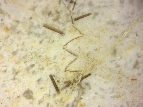
presence of mineral platelets, probably baryte crystals, arranged
near the conspicuous line with several kinks can mislead to the
suspicion of a hidden reason behind it but is
purely incidental here (Fig.1).
Simply ignoring the platelets may be a good guess to start with. The
thin dark line is really a former narrow crack traversing the partially
solidified outer aerial root (Fig.2). Since cracks tend to move
straight
ahead but never
take a kinky path, there must have been hidden processes at work.
Apparently
the silicification process was still going on after crack formation so
that the gap became filled with silica gel which hardened into
chalcedony while the partially silicified aerial root as a whole had
still retained some ductility. While the aerial roots became gradually
compressed by the overlying sediment, the incompressible crack fill
responded with the formation of kinks near the middle of the root where
the whitish matter apparently had still been less hard.
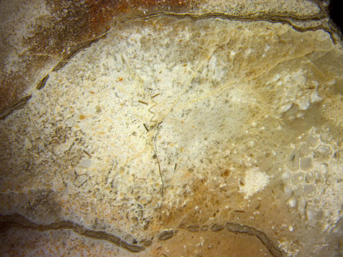
Fig.1: Peculiar structure inside a Permian fossil but unrelated to
palaeobotany. Image width 2.2mm.
Fig.2: Outer aerial root of the tree fern Scolecopteris,
slightly compressed, with kinked crack fill seen as a thin dark line.
Image
width 11mm.
Closer
consideration reveals that the phenomenon of kink formation is fraught
with problems of continuum mechanics. The crack now seen in
cross-section as a thin line must have formed in a soft elastic solid,
probably
silica gel. For reasons unknown, the silica-rich water in the gap
turned into gel and brittle siliceous matter while
the surrounding gel still retained some degree of ductility. This
follows from the absence of secondary cracks beside the kinks. Hence,
the kinked line in Figs.1,2, which is really a section of a
kinked laminar crack fill, is the non-trivial
outcome of the common action of
elasticity and ductility.
By leaving mechanics aside
and turning to palaeobotany again, a better preserved root from the
same sample is shown in Fig.3.
Fig.3: Outer aerial root of the tree fern Scolecopteris with
aerenchyma. Image width
11mm.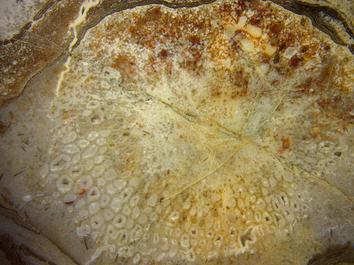
Unlike the particular crack in Figs.1,2, the "normal" cracks
in Fig.3
were formed when all was hard and brittle and thus are uninteresting.
The huge aerenchyma cells with diameters up to 0.5mm are supposed to
have been filled with air and thus have kept the tree afloat. Many are
seen well preserved here but the comparatively tiny tracheids in the
central strand seem to be all collapsed.
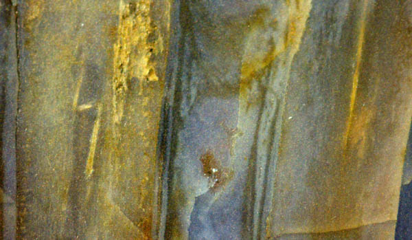 Less wondrous than the kinked hard sheet in
Permian chert (Fig.1) is the kinked
stiff rod in Devonian chert (Fig.4), included here for
comparison. It
is the xylem strand of Rhynia
acting like a laterally confined rod unable to
bear an increasing
load. While most of the
plant responded by gradual deformation, the
stiff silicified xylem became mechanically unstable and suddenly
relieved the
compressive stress by a sideward jerk making the two kinks seen in
Fig.4 on the left.
Less wondrous than the kinked hard sheet in
Permian chert (Fig.1) is the kinked
stiff rod in Devonian chert (Fig.4), included here for
comparison. It
is the xylem strand of Rhynia
acting like a laterally confined rod unable to
bear an increasing
load. While most of the
plant responded by gradual deformation, the
stiff silicified xylem became mechanically unstable and suddenly
relieved the
compressive stress by a sideward jerk making the two kinks seen in
Fig.4 on the left.
Fig.4: Lengthwise cut of two Rhynia,
with and without kinked xylem strand.
Image width 5mm.
Samples: Bu13/35 (0.15kg) Parts1,2, found at the
classical Scolecopteris-
site, Doehlen Basin, in 1999: Figs.1-3. Rh4/57
(0.51kg) Part2, found near Rhynie in 2009: Fig.4.
H.-J. Weiss
2021
|
 |
 31 31 |