Rhynie chert sample with peculiar details
An incidental combination of more or
less uncommon features make
this sample worth being commented on: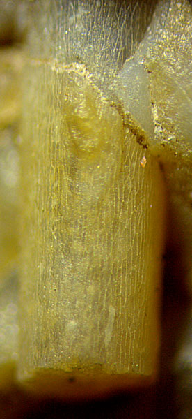
- no other plant than Rhynia seen on the
surface and on cut faces,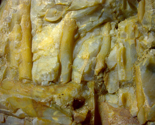
-
except for a 1.5cm bottom layer, Rhynia
in upright
growth position,
- kinks indicating differential
silicification rates,
- mixed
calcite and quartz in former cavities,
- first (?) evidence of violet calcite (?) in
Rhynie chert,
- uncommon ellipsoidal
chlamydospores of some fungus.
Several
upright shoots of Rhynia
and half pipes left by shoots that must be on the other side of the
divide now are seen in Fig.1. They are not
seen in full height in this chert layer of about 9cm thickness.
Possibly their upper parts had been above the water level, thus did not
become silicified but decayed and disappeared. (Sample: 0.5kg, found
near Smithston in 2009.)
Often the shoots
laid bare on the outside of the sample by fragmentation of the layer
show the tissue structure of the epidermis, as seen in Fig.2 taken at
another site on this sample.
Fig.1: Sample surface with several upright Rhynia shoots and
impressions. Image width 16mm.
Fig.2 (far right): Rhynia
shoot, 1.5mm wide, with epidermis
pattern and one of the typical warts (below the crack).
Fig.3 (below): Two Rhynia
shoots on a cut face, surface deformed by stiff
layer kinked under load from above: outward
kink (below left), inward kink (above right); former voids with clear
calcite, quartz, and bluish chalcedony. Image
width 14mm.
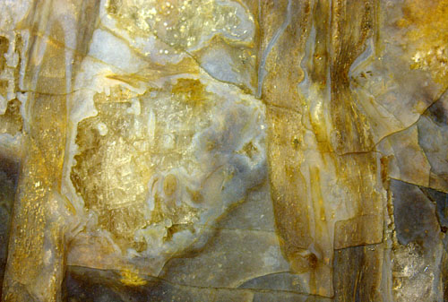
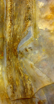
Fig.4 (left): Inward kink, detail of Fig.3.
The deformations on the surface of the shoots (Figs.3,4,5)
indicate the
transient presence of a stiff tube around the shoot, surrounded by soft
matter, which is a hitherto unnoticed stage of the silicification
process.
There is abundant evidence in the Rhynie
chert that most often the surrounding water had turned into silica gel
before the plants rotted and shrunk away from the gel. Now it
appears that, at least in this case, silica gel formed first as a layer
on the surface of the plants. This layer became rather stiff while the
surrounding substance remained still soft or even fluid. With this
assumption, the kinks can be understood as the result of mechanical
instability of the stiff tube under the load of
sediment accumulated above in the meantime. This would be
compatible with the established notion of changing currents loaded with
mud during the formation of the Rhynie chert.
Resistance against
lengthwise compression can also be expected from the xylem strand,
which is
known to retain its stiffness while the surrounding tisse decays so
that eventually
it reacts with kink formation (Fig.6).
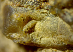
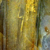
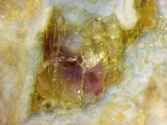
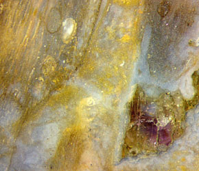
Fig.5 (far left): Rhynia,
2mm, indented
by fragments
of
crushed stiff layer while still soft ?
Fig.6 (left): Rhynia,
2mm, with
kinked xylem strand.
Fig.7 (right): Ellipsoidal fungus
chlamydospores
inside Rhynia;
violet
crystal. Image width 3mm.
Fig.8 (far
right): Violet calcite
(?), detail of Fig.7.
As another peculiarity, fungus
chlamydospores or resting spores with ellipsoidal shape are seen in
degraded Rhynia tissue
in Fig.7. This is worth mentioning since the chlamydospores preserved
in the Rhynie chert, which are often abundant and may be of largely
differing sizes depending on the fungus species,
are nearly always spherical.
Numerous
former cavities in the chert, doubtless filled with water at an early
stage of chert formation, are now seen on the cut faces as crystalline
inclusions consisting of a random assemblage of quartz and calcite
(Fig.3). At the sample surface, no calcite is left, obviously dissolved
since this layer fragment had been exposed to weathering, possibly
since glacial times. The cavities seen in Fig.1 (below and at the edge
on the right) contain only quartz crystals but no calcite.
Most
unusual are the violet spots (Fig.8) extending into the depth of the
crystalline inclusions. The colour strongly resembles that of amethyst
but the aspect of the crystalline substance suggests that it may be a
rare violet variety of calcite.
Again it has appeared that various
information may be derived from only one sample, in this case covering
biology (plant growth and anatomic details, fungus organs), mechanics
(kink formation), chemistry (differential silicification
rates),
mineralogy (clear and coloured crystals grown in water).
H.-J.
Weiss 2017
 |
 |
114 |