Damage control in an early land plant
 There are a few causes of damage in
early land plant tissues. The tissue between epidermis and
central strand, called cortex, could have partially decayed in the
living
plant, similar as with hollow trees. Elusive herbivores gnawed holes
into the shoots
or sporangia
to eat the content. Holes in the tissue of
living plants may even arise
not by partial decay of tissue but by pushing it aside. As a rare
coincidence,
these two types of cavities are seen together in one cross-section.
(See
Rhynie
Chert News 117,
Fig.6.)
There are a few causes of damage in
early land plant tissues. The tissue between epidermis and
central strand, called cortex, could have partially decayed in the
living
plant, similar as with hollow trees. Elusive herbivores gnawed holes
into the shoots
or sporangia
to eat the content. Holes in the tissue of
living plants may even arise
not by partial decay of tissue but by pushing it aside. As a rare
coincidence,
these two types of cavities are seen together in one cross-section.
(See
Rhynie
Chert News 117,
Fig.6.)
Some early land plants had already developed precautions against
herbivore attack: bristles on the outside, tubes with deterrent
liquid (?) along the epidermis, or a protective
wall (?) in the cortex.
The
response of the plant to herbivore attack can be more or less intense.
Obviously, the response had been more intense with the early land plant
in
Fig.1 where big cells had grown around the gnawed hole.
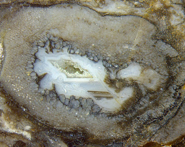
Fig.1: Cross-section of Aglaophyton
with a big cavity due to some herbivore, big cells around, a
few dimly seen sections of coated fungus hyphae grown later in the
water-filled cavity, levels
of watery suspensions, agate-like lining and quartz crystals finally
grown in
the cavity of reducred size. Width of the picture 6mm.
Fig.2 (below): Cross-sections of Aglaophyton with "hollow
straw" aspect, upper one with access hole
in the wall and adjoining tunnel
with surrounding tissue made rot-resistant,
other cortex tissue subsequently vanished owing to rot,
resulting cavities clad with agate-like
linings; lower hollow straw squeezed flat, apparently with similar
damage as above, incidentally
touching the upper one. Width of the picture
7mm. 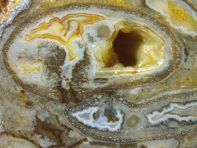
The confusing structure in Fig.2 reveals some response of
the living plant to herbivore attack. Apparently the vicinity of the
tunnel eaten into the plant had been made rot resistant to
avoid the spreading of rot through the tissue. This
is similar as with Fig.1 where the newly grown big cells seem to have
precluded early rotting.
While most of the tissue in Fig.2
decayed, the resistant vicinity of the tunnel
remained as an odd tube and became silicified later.
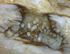 Cell
growth for the repair of the boundaries of a hole is seen with slightly
higher magnification in Fig.3.
Cell
growth for the repair of the boundaries of a hole is seen with slightly
higher magnification in Fig.3.
Fig.3 (right): Cell growth with the aim to fill a hole. Width
2mm.
As a confusing and not yet explained complication, Aglaophyton may or
may not have got a resistant peripheral layer of
cortex tissue (Fig.2) below the often inconspicuous or decayed epidermis. The dark or black appearance of the walls of
the resistant cells seems to be a secondary phenomenon, perhaps a microbial cladding which can chip off.
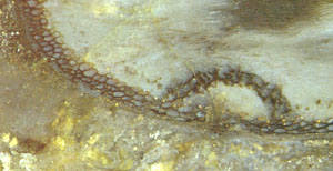
Fig.4
(below right): Hole in the rot-resistant
peripheral layer repaired with a rot-resistant dome-shaped cap of 2mm width.
A peculiar case of damage control is shown in
Rhynie
Chert News 60,
Fig.4, which has been reproduced here (Fig.4). This dome-shaped cap
must have been formed when the now vanished cortex tissue had still been
there and living since it consists of cortex cells made rot-resistant. This
allows conclusions to be drawn concerning the "hollow straw"
phenomenon. It had been explained as a mere diffusion effect in the
sense that dissolved silica entering from outside into a limited depth
preserved only a narrow seam of tissue [1]. That explanation has
already been rejected.
Figs.2,4
suggest another scenario: The plant made a peripheral seam of modified
cortex tissue, perhaps as a protection of the tissue below. This seam
is rot-resistant, thus conspicuous in
advanced stages of decay when most of the cortex tissue is no more there. This
protective seam had got a hole for reasons unknown. The plant covered
this potentially dangerous
hole
with a cap of elegant design. Hence, the plant had been able to respond
to impending danger or attack in flexible ways, which is by
modification of cortex tissue into special layers (Figs.2,4) or by
activation of cell growth (Figs.1,3).
The term "hollow straw" used in connection with
Aglaophyton preserved as a persistent tube might
suggest the idea of retaining some strength while most of the tissue being
decayed. A small hole in the
wall of
the straw would not much affect strength
so that elaborate repair as in Fig.4 suggests a different explanation: The
persistent tubes, in Aglaophyton peripheral only but in Ventarura
always amidst the cortex tissue, are less for
strength than for protection. They possibly delayed the decay. It
remains obscure whether tissue decay in Figs.2-4 proceeded in the live
or dead plant.
Samples: Fig.1: Rh6/9.2 (2002), Fig.2: Rh15/82.4 (2014 obtained from Barron), Fig.3: Rh2/68.1 (2002 obtained from J. Shanks), Fig.4: Rh12/162.2 (2007).
H.-J.
Weiss 2017
[1] C.L. Powell, N.H. Trewin,
D. Edwards: Palaeoecology and plant succession in a
borehole through the Rhynie cherts, ...
Geological Society, London,
Special Publications 180 (2000), 439-457.
 |
 |
118 |