Nothia
sporangia and their peculiar wall structure
The early land plant Nothia
aphylla
found in the Rhynie chert below the hill with the obscure name Tap o'
Noth shows a unique structural component:
aligned tube-like cavities called "giant cells" in [1], where it is
claimed "that they are true cells". Observations [2]
have
led
to the conclusion that the big tubes most probably are of a more
complex
origin involving the dissolution of numerous cell walls. Independent of
this feature it seems appropriate here to draw attention to the fact
that the spore capsules of Nothia
are seen in a variety of shapes on the surface of the chert samples and
on cut faces, hence it may be difficult to tell them apart from
capsules of Horneophyton
and Asteroxylon
which are often found together with Nothia in the same
sample. (Sporangia of Asteroxylon
are
rare.) While the more abundant spindle-shaped sporangia of Aglaophyton
are easily recognized by their mostly elliptical or circular sections,
the irregularly shaped more or less bulky bags which are the Nothia sporangia
may provide odd-shaped sections beside occasional circular
ones.
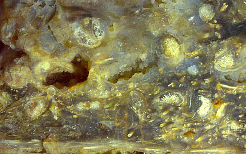
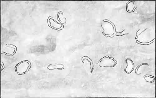
Xylem strands and sporangia with spores are
obviously more resistant to rot than the soft tissue of the plant so
that they are closely stacked in some places in the chert.
Fig.1 with drawing: Nothia
sporangia and xylem strands on an old fracture face of a
chert layer smoothed by weathering. 14 sporangia are visible on this
area of less than 2cm2.
Note
also the numerous persistent xylem strands below.
Apparently the shapes and sizes of the sporangia vary within
wide bounds, which is obvious from
the three pictures below, with equal scale. Some are nearly globular,
others are flat like the uncommonly wide one in Fig.3.
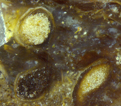
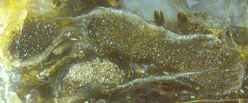
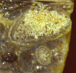
Figs. 2,3,4:
Width 4.8mm, 6mm, 3mm.
Nothia sporangia with spores
varying between black
(below left in
Fig.2)
and completely bleached.
Note the coherent bag of 6mm width
in
Fig.3.
Beside the variability of the capsules, the tube-like
cavities in their walls are worth looking at closely. Although the
sporangia seen in the chert can be numerous, the tubes in the wall are
seldom preserved such that they are clearly visible, and if preserved
well, they are not well seen when tilted with respect to the cut plane.
Hence, the below pictures taken with incident light on arbitrarily cut
faces have been brought about by lucky coincidences.
Fig.5 (left): Three cross-sections of wide tubes below the surface of a
Nothia
sporangium, with pale fill.
Fig.6: Two sporangia of Nothia,
the left one being in the depth and thus seen
from outside.
Fig.7: Detail of the capsule wall in Fig.6: wide tubes hidden behind
the long narrow strips between the cell files.
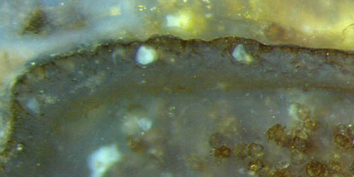
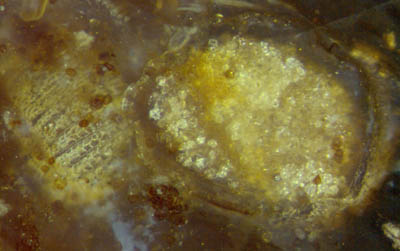
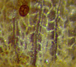
Widths of Figs.5,6,7:
1.4mm, 2.5mm, 0.38mm.
The rare case of contrast by the white chalcedony fill of two
wide
tubes in Fig.5 shows that
the tubes are essentially enclosed within the sporangium wall. A narrow
ridge extends towards the surface where it appears, when looked at
from outside, as a narrow long
strip, about 10Ám wide, as seen between cell files in Fig.7. The tubes
themselves are
about 100Ám across in Fig.5. (According to [1], they can be
up to 1.6mm
long and 0.2mm across. Hence the volume of one tube equals that of
hundreds of normal cells of the capsule wall.) There is at
least one more cross-section of a big tube in Fig.5,
vaguely seen on the left, with a pale bluish spot
inside. (The bigger white spots inside the
capsule are not relevant.)
The epidermis cells arrange themselves in files along the
strips, which
makes the "textile" aspect of the capsule in Figs.6 left and in Fig.7.
(The brown "egg" in
Fig.7 is a separate spore incidentally placed in front of the capsule.)
From the complex structure of the capsule wall as seen here and in
[2] it can be concluded that the evolution of Nothia involved quite a
number of developmental steps, which distinguish it from other "early
land plants" and suggest a closer relationship with the more advanced
zosterophylls, which has been suspected [1] but apparently never
confirmed.
As a well-known fact, tiny contaminations in the silica gel
can largely affect the aspect of the chalcedony owing to
differential silica grain sizes formed in silicification. Hence, the
white fill in some of the tubes possibly indicates a content which had
been chemically different from the surrounding tissue before
silicification, which would
be compatible with the idea of tubes releasing poison or glue if tapped
by creatures trying to get at the tasty spores. This idea differs
essentially from
others proposed in [1], including "large water reservoirs".
Since the surface
tension
of liquids is a dangerous force for small creatures, an earlier
statement [2] may be repeated here: "If tapped by a spore
eater,
a tube under
pressure would spill its content profusely and soil the biting creature
with sticky liquid with possibly fatal effect."
All pictures have been taken from one chert sample of about 0.25kg found in 2003.
H.-J. Weiss
2013,
emended 2014
[1] H. Kerp, H.
Hass, V. Mosbrugger: New data
on Nothia aphylla,
in: P.G. Gensel,
D. Edwards (eds.): Plants Invade the Land, N.Y. 2001.
[2] Rhynie
Chert News 33
 |
 |
57 |


