Asteroxylon sporangia
of questionable shape
Among the 7 “higher” plants found in the Rhynie chert, Asteroxylon is the
tallest and most
advanced one. According to detailed reconstructions pictured in
palaeobotany monographs and on websites, with broad sporangia laterally
attached to the upright axis by means of a short stalk, it
conspicuously resembles some recent clubmoss (Fig.1).


Fig.1: Reconstruction of Asteroxylon
as usually pictured: drawing in [1] after a wax model by Bhutta [2], apparently
embellished to resemble a recent
clubmoss.
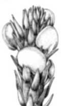
Fig.2, far right: Longitudinal section of Asteroxylon with
two sporangia cut lengthwise, drawn after photograph in [3].
While the sporangia of Aglaophyton and Horneophyton
are not rare and easily
recognized in the chert, those of Asteroxylon are
rather elusive. A photograph by Lyon [3]
is possibly the only picture clearly showing both the lateral
attachment and longitudinal section (Fig.2). Chert samples containing Asteroxylon
with sporangia like those collected by Lyon and
provided to Bhutta
[2] for study are rare.
Doubts concerning the reconstruction in Fig.1
arise from its tortuous way of making: According to [1] it has been
drawn after a wax model kept at the National Museum of Scotland,
Edinburgh. The wax model had been crafted by Bhutta after a series of images of peels as an essential part of his thesis
[2], where it is reproduced in several photographs which do not as much
resemble a recent clubmoss as Fig.1 does.
Thus it appears that Fig.1 is an idealized reconstruction adapted to a
recent clubmoss. According to [2], the sporangia are
“somewhat reniform" and "may show varying degrees of distortion", which
indicates greater variability than suggested by Fig.1.
Own examinations have produced several
small round sections of sporangia near Asteroxylon
axes on cut faces of chert samples but no broad sections
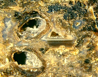
which should be expected from sporangia shaped as those in Fig.1. The
interpretation of sporangia found quite close to Asteroxylon
xylem as in Fig.3 is not easy because what is
seen close together in the chert does not necessarily belong together. Asteroxylon
is often found next to the smaller plant Nothia
in the same sample, and sections of
Nothia
sporangia resemble those in Fig.3. Until Nothia had been
recognized by Lyon
as another species in 1964 [3], it was mistaken for Asteroxylon
branches. Since the two species are
most often preserved in a damaged state, with the
more durable xylem strands and capsules displaced, uncertainty and
confusion seem inevitable.

Fig.3: Sections of two sporangia, or two
lobes of one, partially filled with quartz crystals, only a few spores
left. Note
the Asteroxylon
xylem cross-section above right, also the level
band indicating a prostrate
position of the axis during silicification. Width
of the picture 5mm.
Differences in other anatomical detail have
raised the question of the possible presence of more than one
Asteroxylon
species in the Rhynie chert [4], which
might (partially) explain the apparent variability of sporangia.
With such state of things it is worth while looking for evidence
incompatible with Fig.1, thereby being cautious
not to mistake the possibly also present sporangia of Nothia for those of
Asteroxylon
as it had been done before.
The mutual arrangement of the two sections in
Fig.3 does not look incidental. The cavities seem to extend into and
possibly converge in the depth. If so, they would belong to a
sporangium
whose lumen is broad below but divided into two tapered lobes above. An
interpretation of Fig.3 as a two-lobed Asteroxylon sporangium
would obviously be incompatible with Fig.1.
It has been the aim of this contribution to draw the
attention to the question of how much Asteroxylon can
deviate from its current reconstruction.
H.-J. Weiss
2013
[1] W.N. Stewart,
G.W. Rothwell:
Paleobotany and the Evolution of Plants, Cambridge 1993.
[2] A.A. Bhutta:
Studies on the Flora of the Rhynie chert, Ph.D. thesis, University of
Wales, Cardiff, 1969.
[3] A.G. Lyon:
Probable fertile region of Asteroxylon
mackiei, Nature 203(1964), 1082-83.
[4] W. Remy, H.
Hass:
Langiophyton mackiei, ... . Addendum. Argumenta Palaeobot. 8(1991),
69-117.
 |
 |
53 |


