Aspects of Devonian microbes
One
cannot deny that there is something mysterious in the fact that
cyanobacteria can aggregate in large numbers and form
shapes serving a higher purpose rather than existing as mere
separate unicells. Thereby they may produce even easthetically
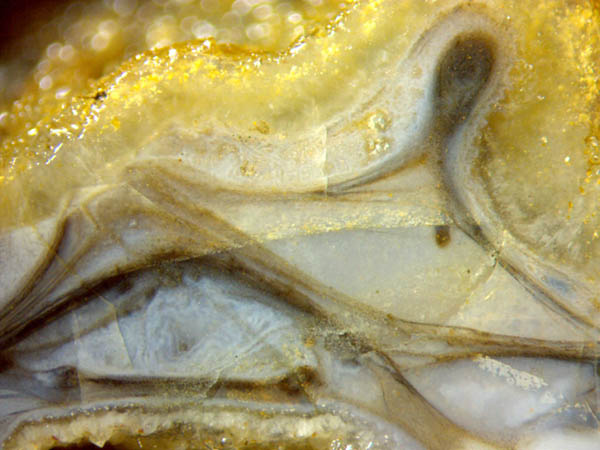
appealing shapes, as seen in Fig.1. Apparently three curved microbial
layers converge to a spot on the left where there had been
another
club-shaped object which had got lost. No
explanation is given here for the formation of the smoothly curved
layers. All pictures have been taken from 6 samples of the Lower
Devonian
Rhynie
chert.
Fig.1 (right): Structure of unknown purpose brought about by
a coordinated action
of a
multitude of unicells. Image width 5.5mm. Figs.1-4 with same
scale.
Fig.2
(below): Similar as with Fig.1. The club-shaped protrusions
are apparently
related
to the big globular object in the centre with multi-layered sheath.
Image
width 6mm.
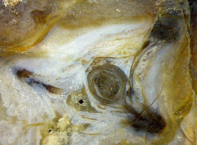
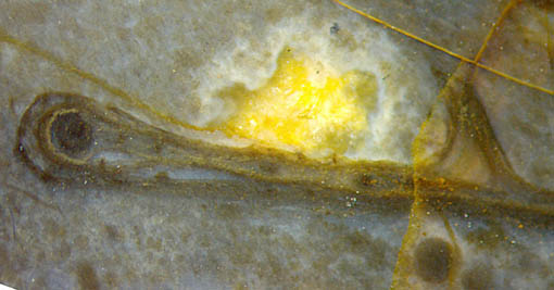
Fig.3:
Clots within a microbial envelope.
Image width 4.7mm.
Fig.4 (below): Horizontal layer with small dark clot below and closed
envelope above obviously made of the same microbes.
Image width 5.5mm.
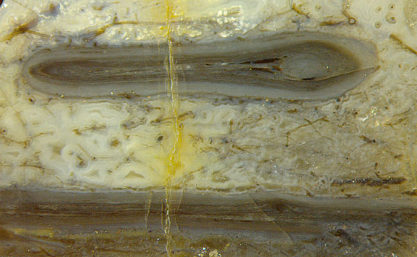
Envelopes
with one or more clots inside have been seen repeatedly but their
formation is still enigmatic. There is no indication that they had been
formed as bulky shapes squeezed flat.
From common features in
Figs.1-4, the presence of (multiple) sheaths forming envelopes around
clots, one may guess that these structures were formed by the same kind
of microbes, or similar ones, which apparently have not yet been
described.
One
can distinguish a few different microbes from the Rhynie chert without
going into details of the cellular structure. For example, Croftalania venusta
[1] is easily recognized if grown in conspicuous tufts of filaments as
in Fig.5 and in Rhynie
Chert News 56, 72.
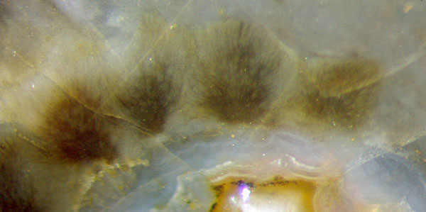
Fig.5 (left): Tufts of the filamentous microbe Croftalania venusta
[1] grown from a temporary silica gel surface towards the
light.
Image width 1.7mm.
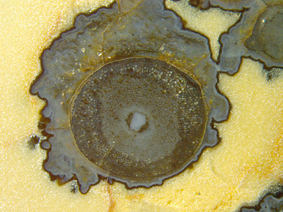
Fig.6 (right): Black coating
on the surface of bluish silica gel around Aglaophyton.
Image width 8.6mm.
Often seen are microbial coatings on plant parts and surfaces
of silica gel. They may have begun yellow but turned brown or black
with time.
From
the aspect of Fig.6 it can be concluded that the silica gel had got a
well-defined boundary before the microbes settled there in a coating of
about 40µm thickness, which makes a black line in cross-section. Higher
magnification reveals that the black line is made up of tiny black
dots. (Same sample as with Rhynie
Chert News 64.)
Apparently
these microbes, too, have not yet been
described.
Another distinctly different kind of microbe is
seen in Rhynie
Chert News 23
as
a unique spherical colony being attacked by
a unique creature, the only specimen ever
seen of a Devonian rotifer.
By an unbelievably lucky coincidence, the polished face only touches
the rotifer and cuts right through the middle of the sphere of about
500 cells, probably cyanobacteria. It is recommended here to look for
more of these elusive spheres and rotifers on Rhynie chert samples
stored in collections so that eventually they become established
components of this Lower Devonian habitat.
The larger microbial formations as seen in Figs.1-6 are not rare in the Rhynie chert.
H.-J.
Weiss
2018
[1] M. Krings, H. Kerp, H. Hass, T.N.
Taylor, N.
Dotzler: A filamentous
cyanobacterium showing structured colonial growth from the Early
Devonian Rhynie chert.
Rev. Palaeobot. Palyn.
146(2007), 265-276.
 |
 |
121 |