Black stains, coatings, and linings in
the Rhynie chert
The Lower Devonian Rhynie chert is famous for its rich detail, mostly due
to fossil plants but also to creatures and mineralisation phenomena.
Some of the structural features are conspicuous owing to enhanced
contrast from black or dark stains, coatings, or linings. Hollow straws
of Aglaophyton
with clearly seen dark cell walls, or what appears to be
such, are rather common
(Fig.1). The hollow straw aspect had been explained as a result of
silicification penetrating into a limited depth [1,2], a
view which has been doubted in [3]. A characteristic tube inside the
rarer
plant Ventarura
(Fig.2), likewise consisting of tissue with
apparently thick and most often dark cell walls, had been regarded as
sclerenchymatic [1]. Contrary evidence has been provided: [4] and Rhynie
Chert News 60,
66.
The
usually inconspicuous cuticle on the epidermis of land plants is
occasionally covered with a pale layer which may turn black and thus make
clearly visible contours of plant sections (Figs.2,3). The cuticle with
(stained) coating can become
detached from the degrading plant and deform under residual
stresses, thereby making curved shapes (Fig.4).
Other curved shapes not related to biological structures arise from
linings
of former swamp gas bubbles which later became filled with water and silicified (Fig.5).
The transition from pale to dark or black
can be smooth or rather sudden (Figs.4,5,7).
Since
there is fossil evidence indicating that the dark appearance is
not merely a stain of the very wall or cuticle matter but is due to
some added layer which can also be pale, a microbial involvement
suggests itself.
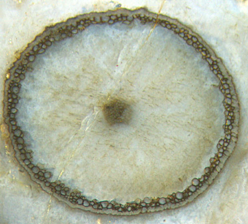
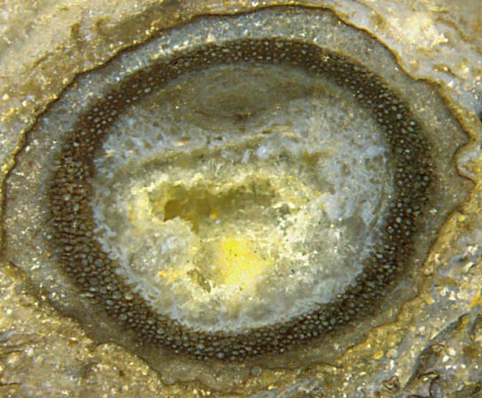
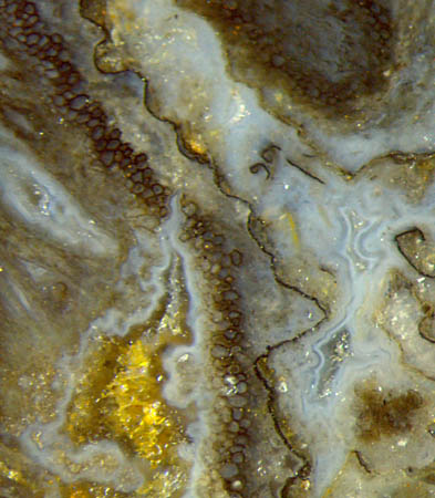
Fig.1: Aglaophyton
"hollow straw", 2.2mm, tissue mostly decayed
except for a black-stained tube
adjacent to the poorly preserved
epidermis.
Fig.2: Ventarura,
characteristic aspect with shrivelled contour and well
preserved hollow cylinder mimicking sclerenchyma, other tissue
decayed. Width of the picture
5.5mm.
Fig.3: Two Ventarura sections:
shrivelled contour with black stain (left) and without
or pale stain (above right). Width of the picture 2.5mm.
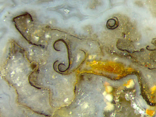
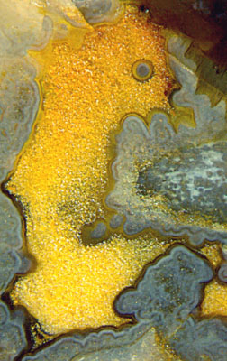
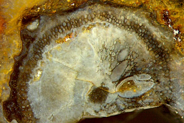
Fig.4 (connected to Fig.3 on
the right, enlarged): Detached fragments of coated cuticle,
deformed by residual stresses, with pale coatings partially stained
black. Note the long continuous patch of cuticle in the lower half of the
picture, extending beyond the
frame below, with the transition between pale and black varying between
smooth and sudden. Note
also the dimly seen crack in the chalzedony below, which chooses the
mechanical discontinuity provided by the cuticle for an easy path.
Width of the picture 1.5mm.
Fig.5: Former
water-filled cavity in silica gel with coatings on the
gel surface, varying between pale and dark, gel later turned into
bluish
chalzedony, cavity later filled with yellow-stained quartz. Height of
the
picture 5.5mm. (See also Rhynie
Chert News 64.)
Fig.6: Ventarura, rare case of abnormally grown inner tissue, preserved and stained dark.
Width of the picture 4.3mm.
The
cross-section in Fig.6 is rather peculiar and deserves a
special comment. The shrivelled circumference, the vanished adjacent
tissue, the dark ring of conspicuously preserved tissue, the vanished
tissue inside, and the central strand are the usually seen features of
Ventarura.
What is
quite unusual with this particular specimen is the cortex tissue on the
right half, grown in a misguided way and being distinctly seen
owing to dark stain. Similar misguided growth,
probably due to the influence of
fungi present in the live plants, is found in several other
species of early land plants, as discussed in Rhynie
Chert News 4,
21,
54.
In the present case the fungus (?) did
not only cause the plant to grow disproportionately big cells which
might
have become voids. Apparently it also made the affected part of the
tissue
decay-resistant so that there was enough time available for a dark
stain to settle on the cell walls before silicification, which makes
the smaller ones of the affected cells resemble those of the
characteristic (dark) ring.
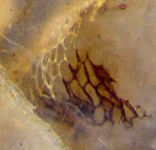 Fig.7: Curved patch of epidermis surface of
unidentified
sporangium with sharp boundary between pale and dark brown. Width
of the picture 0.65mm.
Fig.7: Curved patch of epidermis surface of
unidentified
sporangium with sharp boundary between pale and dark brown. Width
of the picture 0.65mm.
Several
observations indicate that dark coatings can become bleached by oxidation of the carbonaceous decay products of
the organic substances so that they look similar as in Fig.7. The
oxygen can enter by diffusion through the
chalzedony. Hence, there is the additional uncertainty that pale coatings can be bleached or originally pale.
The observations illustrated here and in Rhynie
Chert News 60, 66, 87 suggest
that the various dark formations, or part of them, may have
something in common.
A few
conclusions may be stated:
- Deposits on cell walls, cuticles,
and former
silica gel surfaces in Rhynie chert are often
of microbial origin.
-
The aspect of the said deposits can vary between pale and black. The deposits may escape notice when overall pale.
-
The said deposits had been formed only on substrates that had
persisted in water long enough.
- Tissue degrading early had not
become coated.
- The rings of Aglaophyton
"hollow straws" and of Ventarura
are due to cell walls made decay-resistent in the
live plant.
-
The said rings are conspicuous because of both their mere persistence
and
the pale or stained cell wall coatings.
- The coating is not strongly bonded to the
substrate but may become reduced to detached flakes.
Perhaps the phenomenon of black stains in Rhynie chert can be approached and finally explained by separating it into
simpler items:
-
How do parts of tissue acquire such decay resistance
that they persist among decaying tissue
and get coated cell walls ?
- Which aquatic microbes form coatings on persistent
substrates ?
- How do the coatings become stained ?
H.-J.
Weiss
2015, revised 2016
[1] C.L.
Powell, N.H. Trewin, D. Edwards: Palaeoecology and plant
succession in a borehole
through the Rhynie cherts, ...
Geological Society, London,
Special Publications 180 (2000), 439-457.
[2] www.abdn.ac.uk/rhynie, Chapter Taphonomy.
[3] A. Channing:
Processes and
Environments of Vascular Plant Silicification: Thesis, Chapter
6, Cardiff University, 2001.
[4] H.-J.
Weiss: Rhynie chert -
Implications of new finds,
European Palaeobotany and
Palynology Conference 2014, Padua.
 |
 |
83 |