Aglaophyton spores
decayed inside sporangia
 Aglaophyton,
the most abundant of the early land plants preserved in the silicified
Early Devonian habitat known as Rhynie chert, still offers interesting
details which occasionally even contradict views established in the
scientific literature. The maximum diameter of its fusiform sporangia
is 4-5mm
according to [1], 4mm
according to [2], but
6.5mm in Rhynie
Chert News 11,
about 7mm in Rhynie
Chert News 61,
and 7mm in Fig.1.
The twisted sporangia had led to the idea that the twist came with the
splitting [1], which has been refuted. These references show that it is
still worthwhile to closely look at Aglaophyton.
Aglaophyton,
the most abundant of the early land plants preserved in the silicified
Early Devonian habitat known as Rhynie chert, still offers interesting
details which occasionally even contradict views established in the
scientific literature. The maximum diameter of its fusiform sporangia
is 4-5mm
according to [1], 4mm
according to [2], but
6.5mm in Rhynie
Chert News 11,
about 7mm in Rhynie
Chert News 61,
and 7mm in Fig.1.
The twisted sporangia had led to the idea that the twist came with the
splitting [1], which has been refuted. These references show that it is
still worthwhile to closely look at Aglaophyton.
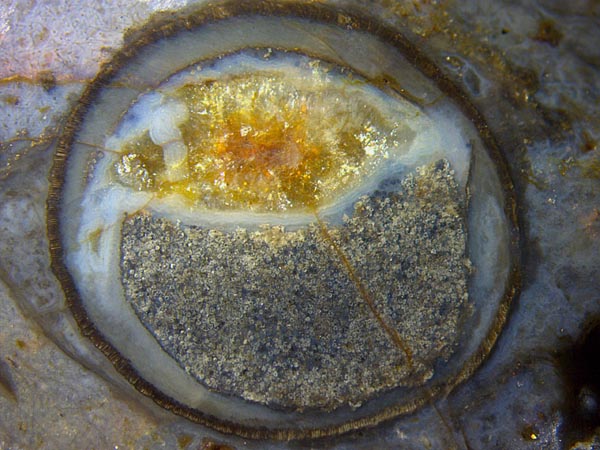
Fig.1: Big Aglaophyton sporangium,
diameter 7mm, filled with damaged spores,
bluish chalcedony, and coarse
crystalline quartz.
The
sporangium seen here is highly peculiar for more than one reason. Its
diameter of 7mm is among the biggest ever seen. It is not obvious why a
well-defined curved boundary had separated the mass of spores below
from a
water-filled cavity above. The cavity walls and
a few fungus hyphae grown in the silica-rich water
(on the left) had become
coated with silica gel turning into bluish chalcedony. Later the
remaining cavity became filled with coarse
crystalline quartz.
Most remarkable is the uncommon aspect of the spores, not chewed up by spore eaters
but severely damaged nevertheless (Figs.2,3). These detail
images are representative for the heap of spores in Fig.1. Since
essentially all spores seem to be affected by the same type of damage,
one may wonder whether decay due to being submersed for a long time
before silicification or a quite different phenomenon had been at
work. This small chert sample (15g) does not offer another
big sporangium for comparison but a smaller sporangium
(Fig.4) provides additional insights.
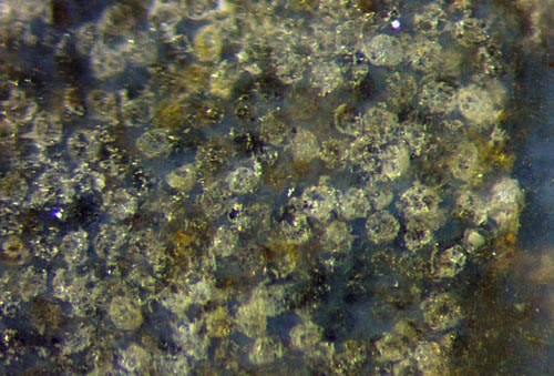
Figs.2,3:
Damaged Aglaophyton
spores
inside the sporangium of Fig.1. Image widths 2mm, 1.2mm.
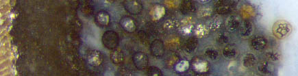
Fig.4 suggests the idea of some destructive process encroaching
on the nearly globular
spores or tetrads, mostly pale and a few black
as seen above left. (The
black ones seem to be coated with a microbial layer.) Below left, only
a few spores are
seen affected, apparently with holes in the wall. Quite different is
the aspect on the right where the destructive
process had reduced the spores to debris.
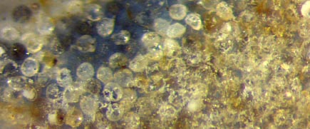
Fig.4 (right): Aglaophyton spores, well preserved or damaged, inside a smaller sporangium,
same sample as Fig.1. Image width 1.1mm.
Fig.5: Detail of an Aglaophyton
sporangium in Rhynie
Chert News 166
with an enigmatic structure
clearly
seen inside a transparent spore. Image width 1.1mm, same scale of
Figs.3-5.
No explanation is proposed here for the
destruction of spores inside sporangia. A related observation (Fig.5),
where
a transparent spore is seen with a definite enigmatic structure inside,
can possibly lead the way to an explanation.
Nearby spores, too, are structured inside but less clearly seen.
Sample: Rh4/67 (15g) found in 2009.
H.-J. Weiss 2021
[1] D.S.
Edwards : Aglaophyton
major, a non-vascular land-plant from the Devonian Rhynie
Chert.
Bot. J. Linn. Soc. 93(1986), 173-204.
[2] abdn.ac.uk Aglaophyton
 |
 |
178 |