Spore clots in Rhynie chert
Mature
spores as in Fig.1 are released from the sporangia as loose grains, as
expected. So it is surprising that spores are not seldom seen in
globular lumps (Figs.2-6).
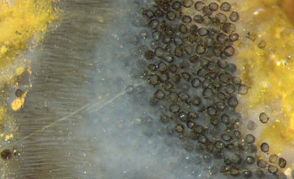
Fig.1 (right): Detail of Aglaophyton
sporangium:
wall with so-called palisade cells (left), sporogenic tissue, spores.
Width of the picture 1.7mm.
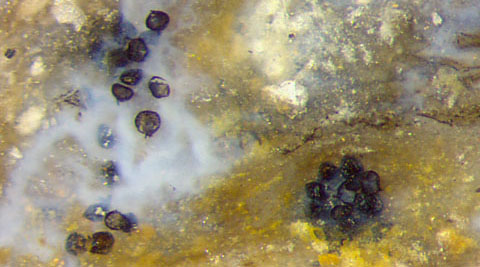
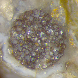
Figs.2,3: Aglaophyton spores,
separate and in globular clots. Height of the pictures 0.8mm. All
pictures of same scale.
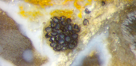
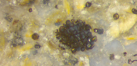
Fig.4 (far left): Aglaophyton
spores
in a globular clot.
Width of the picture 1.4mm.
Fig.5 (left): Aglaophyton
spores
in a globular clot, apparently partially chewed up.
Width of the picture 1.4mm.
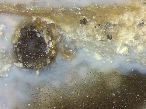
Fig.6 (right):
Aglaophyton spores
in a globular clot, with mineral debris apparently glued to the surface.
(One circular spore contour is faintly seen above left.)
Width of the picture 1.4mm.
The peculiar clots shown here require an
explanation.
The statement in [1] that "evidence for deliberately targeted spore
feeding in the Early Devonian is not conclusive" had been contradicted
by own
observations in 2005: There were spore eaters around when the early
land plants became preserved in the Rhynie chert. This has suggested
a phantastic idea: Possibly some creature collected the scattered
spores and glued them into spheres for transport,
storage, and later use as food. A few
spores nibbled off the surface of the clot in Fig.5 seems to support
this assumption. The mineral debris sticking to the clot
in Fig.6 supports the idea of glueing.
Unrelated
to the clot problem but worth mentioning are lots of tiny black dots in
Fig.6, probably microbes stuck to a former silica gel surface.
Annotation
2021: An uninspired explanation for the existence of spore clots is
offered in [2]: "Clusters of spores or spore balls are typically found
in the chert matrix, suggesting that the spores may have been shed en
masse (Fig.8.31)." (The scale bar in that figure should be rather
0.1mm than 1mm.)
A sphere with a
thousand spores like the one in Fig.3 cannot simply result from
shedding. Also it cannot have been formed inside the sporangium since
mature
Aglaophyton
sporangia are known to split open with a longitudinal
fissure too narrow
for clots.
Samples:
Rh9/93 (0.55kg), found by S. Weiss in
2011, cut into 10 parts, Part5: Fig.1.
Rh2/4, obtained from Shanks in
1998, cut into 4 parts, Part2 returned. Part1 (slab, given to J. Gardavsky):
Figs.5,6; Part3 (slab, in the own
collection): Fig.2,4.
Rh4/22 (1.3kg), found by S. Weiss in
2000, cut into 2 parts, Part2: Fig.3.
H.-J.
Weiss
2019, 2021
[1]
K.S. Habgood, H. Hass, H. Kerp:
Evidence for an early terrestrial food web: coprolites from the Early
Devonian Rhynie chert.
Proc.
Roy. Soc. Edinburgh, Earth Sci. 94 (2004), 371-389.
[2] T.N.
Taylor,
E.L. Taylor, M. Krings: Paleobotany,
Elsevier 2009, Fig.8.31.
 |
 |
149 |