Nematophyte tubes and "branch knots"
Among
the questions arising in connection with the enigmatic nematophytes,
two of them seem to be most intriguing: Where do the tube-like
filaments come from, and what is hidden inside the lumps known as
branch-knots
? It has been tempting to answer
the two questions by reducing them to one phenomenon: The tubes have
been supposed to branch profusely [2] inside the branch-knots,
then come out. The idea that the tubes seen outside the knots are also
present within has been supported by the
questionable statement in [1], p.80, that "tubes can be seen entering
and leaving them.".
It is emphasized here that there is evidence from
several nematophytes
that the clots called branch-knots are not mere dense
tangles of tubes, branching or not, but objects of their own. They can
be so small
that there would not be enough space inside
for "very
tightly coiled ... tubes showing repeated and closely spaced branching"
[3], as seen in Rhynie
Chert News 136,
there Fig.3.
The big clots of the hitherto unknown nematophyte in Rhynie
Chert News 92
are distinguished by a definite boundary and an apparently mainly
structureless
interior. The absence of tubes emerging radially from the clots
enables the tubes to form a parallel texture. Probably they emerge from
the clots in such a way that they fit into the texture immediately. 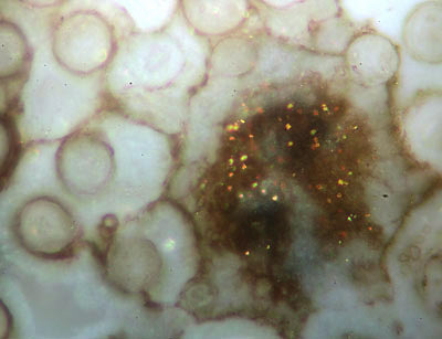 A smaller clot among the big ones in Rhynie
Chert News 92
, there on Fig.2, seems to be different: Tiny reflecting crystals,
perhaps pyrite, indicate a deviating chemical composition, possibly due
to another stage of growth at the time of silicification: here Fig.1.
The vaguely seen undefined structure inside the clot
could possibly provide the expected but elusive connection between
interior and surroundings. Shrinkage
cracks in the gel outside the clot may mislead to the illusion
of cell walls, which is not relevant here.
A smaller clot among the big ones in Rhynie
Chert News 92
, there on Fig.2, seems to be different: Tiny reflecting crystals,
perhaps pyrite, indicate a deviating chemical composition, possibly due
to another stage of growth at the time of silicification: here Fig.1.
The vaguely seen undefined structure inside the clot
could possibly provide the expected but elusive connection between
interior and surroundings. Shrinkage
cracks in the gel outside the clot may mislead to the illusion
of cell walls, which is not relevant here.
Fig.1: Small clot with vaguely seen structure of obscure significance,
surrounded with nematophyte tube cross-sections up to 60µm.
Image width 0.4mm. Sample: Rh2/81.3.
Obviously, the big tubes
in Fig.1 are unlikely to branch within the small clot but the hardly
seen much smaller tubes might do so.
Unexpected
support for the idea of "normal" tubes being somehow connected to small
branching tubes inside clots comes from another unknown nematophyte (in
Rhynie
Chert News 13
, 156
,
there Fig.4), which does not grow lots of clots with definite outline
as the one in
Rhynie
Chert News 92
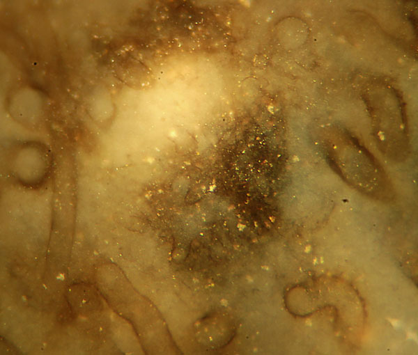
and Fig.1. The clot in Fig.2 is the only one seen
in this specimen, and it has not got any visible
boundary but a strangely shaped object inside which could possibly
connect the poorly seen
thin branches inside to the big tubes outside.
Fig.2: Nematophyte clot with branching object and
small tube parts inside, surrounded by
nematophyte tubes with cross-sections up to 60µm.
Image width 0.6mm. 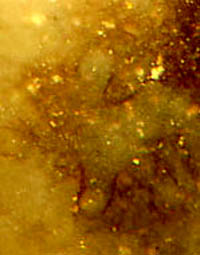 Sample:
Rh13/7.1.
Sample:
Rh13/7.1.
Fig.3, detail of Fig.2: Connecting piece (?). Image
width 0.15mm.
Figs.2,3
are compatible with the less conventional interpretation that the
60µm-tubes seen outside do not
emerge from inside the clots but are formed on their surface by fusion
of smaller tubes: about 20-25µm with this specimen.
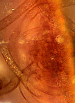
Fig.4 (right): Small Nematoplexus
clot with the onset of an 11µm-tube. Image width 0.1mm.
Sample: Rh15/79.4.
The phenomenon of tube formation from two or more smaller
ones is apparently rather common. It has been
observed recently, on a smaller scale, with Nematoplexus: Fig.4,
which is a
detail of Fig.2 in Rhynie
Chert News 134,
shows one of the rare occasions where a tube is seen
being formed near the surface of a clot. Here, the onset of an 11µm-tube is made up of two very short converging parts, 6µm
and 8µm wide, possibly connecting the tube with some internal structure. There are no clearly seen tubes inside the clot.
Note the largely differing tube sizes among the
nematophytes:
up to 60µm and more in the undescribed species
(Figs.1-3), and mostly 7-15µm in Nematoplexus.
Samples: All photographs taken by Gerd Schmahl, Dresden.
Fig.1: Rh2/81 (0.63kg), obtained from Shanks in
2003, Part 3.
Figs.2,3: Rh13/7 (0.25kg), found by S.
Weiss in 2005, Part 1.
Fig.4: Rh15/79 (0.27kg), obtained from Barron in
2014, Part 4.
H.-J.
Weiss 2020
[1] A.G. Lyon:
On the
fragmentary remains of an organism referable to the nematophytales,
from the Rhynie chert, Nematoplexus
rhyniensis.
Trans. Roy. Soc. Edinburgh
65(1961-62), 79-87, 2 tables.
[2] T.N. Taylor
et al.: Paleobotany. Elsevier 2009, p.180.
[3] www.abdn.ac.uk/rhynie/nemato.htm
 |
 |
158 |