Multiple
xylem strands in an unidentified Lower Permian plant from Döhlen basin
Multiple xylem strands are known from the stems
of fossil trees: Ribbon-like strands of primary xylem run along the
center of the Psaronius
tree ferns, their number increasing as the center becomes wider with
height [1,2]. Wooden strands of various shapes are present in the stem
of medullosean seed ferns and in Rhexoxylon,
an Upper Triassic /Lower Jurassic tree of uncertain affiliation. Thin
multiple strands have been found in several paleozoic seed fern frond
stalks [3]. Double xylem strands are a common
sight in the Lower Devonian Rhynie chert, where the prongs of the
forking central strand of Nothia
keep growing closely together for some distance while the
shoot
does not yet fork. Even four-fold strands can form
in this way, or three-fold ones if the second forking is not
simultaneous in the two prongs. A peculiar type of unsymmetric growth
after forking
of
the strand is observed with Trichopherophyton.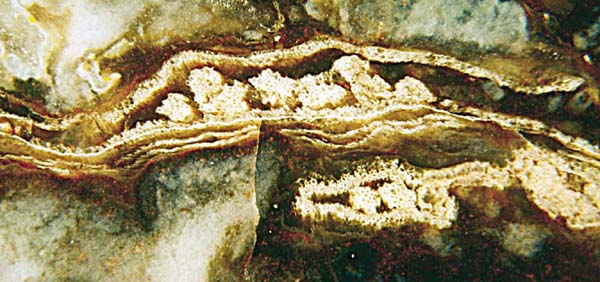
Fig.1: Multiple xylem strands on a Lower Permian plant cross
section.
Width of the picture 7mm.
Apparently the xylem strands found in one chert sample from Döhlen
basin (Fig.1) differ from any one of the various types of multiple
strands mentioned above.
As it is often observed with xylem strands, they are much better
preserved than the surrounding tissue. The latter has mostly vanished
in the present sample. What has remained in addition to the strands is
a well preserved envelope, probably a kind of sclerotic sheath, and
a faintly seen epidermis with cells 25 to 40 µm wide. There had been
some tissue between the sheath and the epidermis, judging from a gap or
squeezed cells seen there in some places. (Two thin-walled squeezed
empty sheaths are seen in Fig.1 below the big sheath with the 8
strands.)
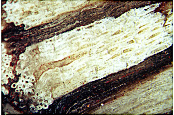
Fig.2: Kinked xylem strand with cells cut both across (far left) and
lengthwise. Note the arrays of pits faintly
seen on some of the cell walls. Width of the picture 1.6mm.
The overall structure of the cross-sections with separate strands of
primary xylem enclosed with a sheath (Fig.1) resembles
that of the Psaronius
center but the pitted cell walls of the tracheids
(Fig.2) and the peculiar cells oriented across the strand do not seem
to be compatible with marattialean ferns. The structure is partially
similar to but also different from that of seed fern frond stalks, as
of Stenomyelon, for example [3].
Detached sporangia
resembling those of leptosporangiate ferns, sterile fern-like pinnules,
and plant debris of uncertain origin contribute to the confusing but
intriguing impression made by this chert sample.
This unique sample had been collected and provided by W. Schwarz (1952-2012).
H.-J. Weiss
2013
[1] G.W. Rothwell, A.H. Blickle:
Psaronius magnificus
..., J. of Paleontology 56(1982), 459-468.
[2] H.
Steur, H. de Kruyk: Psaronius, een boomvaren ... (in
Dutch), Grondboor & Hamer nr. 3/4 (2004), 75-83.
[3] T.N.
Taylor, E.L. Taylor, M. Krings: Paleobotany, Acad. Press
2009
|
 |
 11 11 |