Zosterophylls with peculiar branching
mode ?
Two less-common plants in the Rhynie chert, Trichopherophyton
and
Ventarura,
have been assigned to the zosterophylls, a group of plants
whose fossil evidence seems to be restricted to the Devonian. The
zosterophylls are of phylogenetic interest as they are
believed to be ancestral to the lycophytes including the recent
clubmosses.
Trichopherophyton
is not really rare in the chert but apparently had
often been overlooked
because the upper part of the plant with its
characteristic spinous hairs is found much less often than the hairless
lower parts which probably were subterraneous or submerged and thus had
a better chance of silicification. The strange phenomenon of one
Trichopherophyton
axis or shoot inside another one (Fig.1) had been
observed before and interpreted as incidental intrusion [1,3]. A quite
different interpretation is proposed here.

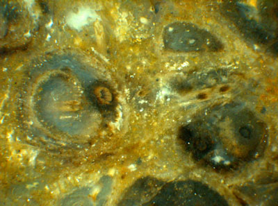
Fig.1: Trichopherophyton
cross-sections: new shoots forming
inside older ones as seen on the raw surface of the chert sample.
Width
of the picture 7.7mm.
Closely spaced Trichopherophyton
cross sections can be found in large
numbers even on faces of small chert samples, which implies that they
are sections of upright axes since creeping axes most probably would
have random directions in a plane. It is surprising that sections of
the one-in-another type are seen rather often. Also it appears that the
allegedly invaded axis is not always in a state of decay, and there are
rare cases of well preserved samples apparently showing coherence of
the tissue across the boundary of the inner axis (Figs.2,3).
These observations suggest a much more interesting
interpretation than
incidental intrusion or other sophisticated
explanations based on unlikely asumptions as in
[1]: After forking of the central strand, the
two
prongs proceed inside the common shoot for a while and move apart until
eventually one of them stakes off a circular claim by surrounding
itself with a distinct borderline within the common tissue.


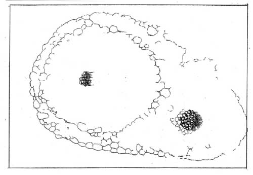
Fig.2 (right): Trichopherophyton
cross-section of composite
type, no indication of squeezed tissue.
Width of the picture
1.9mm.
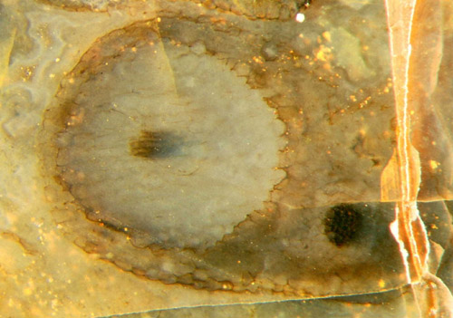
Fig.3: Same as Fig.2. The apparent coherence of
tissue across the inner boundary seems to rule out the interpretation
favoured in [1,2] as
one axis intruding into another one.
As seen in
Figs.2,3, this partitioning of the cross-section goes without
squeezing. Apparently the inner axis is suordinate at first, as
suggested by the smaller diameter of its central strand (Figs.1,2), but
further on it seems to take the lead while the outer part becomes
narrower and eventually wilts (Fig.4). The process may repeat itself so
that finally one or more shoots look like being wrapped into old
sheaths.
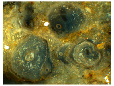
 Fig.4: Same as Fig.1, advanced
stages of new shoots forming inside older ones, with the inner shoot
dominating (below left) and the outer part being reduced to a
sheath which finally may split so that a crescent-shaped
residue of the old shoot is left (below centre)
which has become detached from the
new shoot. Width of the picture 6.5mm.
There is at least one benefit of such growth mode: Old plant parts
covering the base of fresh shoots is a means of multiple protection
well known from flowering plants, as onions, for example. It would be
interesting to know whether this peculiar way of growing protective
sheaths as suggested by the aspect of Trichopherophyton
cross-sections
is unique and restricted to the zosterophylls or found in other plants,
too.
Since there seem to be good reasons for one axis growing inside another
one, it is not surprising that this phenomenon has also been found with
the other zosterophyll in the chert, Ventarura. A few
cases from
Windyfield chert, which is a separate variety of Rhynie chert, are
mentioned in [2], and one is shown there and interpreted as a rhizome
growing along a decaying fragment, despite of its vertical orientation.
Own finds suggest that the lower part of Ventarura often
branches in
the same peculiar way as Trichopherophyton
does so that there is no
need to invoke improbable incidences for an explanation.
It has to be mentioned that the above tentative interpretations are
based on sections seen on the sample surface and on arbitrarily chosen
cut planes. Serial sections could possibly provide more insight and
lead to better substantiated conclusions.
H.-J. Weiss
2006
[1] A.G.Lyon,
D.Edwards: The first zosterophyll from the
Lower Devonian Rhynie Chert,
Trans. Roy. Soc.
Edinburgh, Earth Sciences, 82(1991), 323-332.
[2] C.L.
Powell, D. Edwards, N.H. Trewin: A new vascular
plant from the Lower Devonian Windyfield chert, Rhynie, NE Scotland.
Trans. Roy. Soc.
Edinburgh, Earth Sci. 90(2000 for 1999), 331-349.
[3] D.
Edwards : Embryophytic sporophytes in the Rhynie and
Windyfield cherts.
Trans. Roy. Soc.
Edinburgh, Earth Sci. 94(2004 for 2003), 397-410.
 |
 |
16 |


 Fig.4: Same as Fig.1, advanced
stages of new shoots forming inside older ones, with the inner shoot
dominating (below left) and the outer part being reduced to a
sheath which finally may split so that a crescent-shaped
residue of the old shoot is left (below centre)
which has become detached from the
new shoot. Width of the picture 6.5mm.
