Maggot fern disputes
Why a Permian tree fern had got the
name "maggot fern", which is Scolecopteris
in Latin, can easily be guessed from the two pictures below,
taken from either half of a cut chert boulder collected at the type
locality of this fern in 1997. A few
samples found before 1800 had aroused the imagination then, and given
rise to interpretations as creatures, until the fossil had been
recognized around 1802 as a plant and described as Scolecopteris elegans
in 1837 [1]. This fossil had considerably contributed to the
development of palaeobotany as a science, hence it is worth looking at
in detail. Later it became obvious that the foliage belongs to the
beautiful silicified Psaronius
tree trunks.
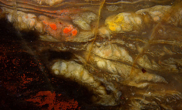

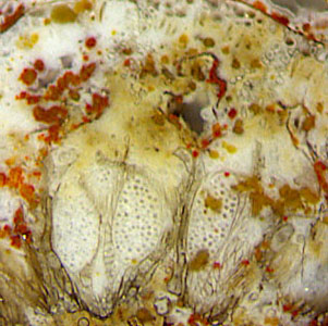
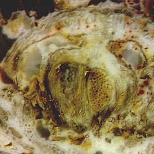
Figs.1,2: Maggot fern (Scolecopteris)
pinnules in various states of preservation, more or less deformed,
suggesting interpretations as grubs or nymphs of
some insect. Images (widths 7mm and 8.5mm, same scale) taken
from the cut halves of one chert layer fragment.
Apparently
some of the capsules in Fig.1 had not been fully grown before the
pinnules fell down into the water, accumulated
there, became deformed, and silicified.
Note the red and yellow stains of spores in the mature capsules in
Fig.2.
Problematic is another
chert sample from the same locality with distinctly seen pinnules on
the surface (Figs.3,4). Plant parts on chert layer surfaces
are clearly seen if they are preserved as hard chert after the
adjoining matter had been eroded away or spalled off.
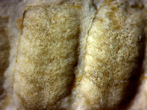
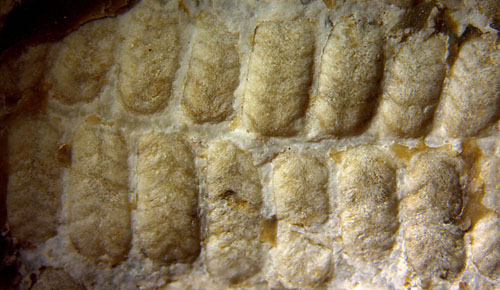
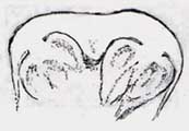
Fig.3 (near left): Maggot fern
pinna
on the raw surface of a chert sample, pinnules
apparently not deformed.
Angle of veins about 60°. Same pinna as in [2],
Fig.8, and [3], Fig.210A. Image width 11mm.
Fig.4 (far left): Pinnules from Fig.3, upper
row; epidermis texture following the veins towards the
margin. Image width 3.5mm.
The pinnules touch laterally in the depth, which is not seen here since
the gaps are filled
with whitish chalcedony. Apparently the fill had been left over when a
crack propagated along the cuticle on the
epidermis, jumped from one pinnule to the next, thus
separating
the
whitish layer and laying bare the pinna as we see it now. The
waxy cuticle seems to have
provided a potential crack path for
easy propagation.
This
fern has been interpreted as Scolecopteris elegans in
[2,3], which is not compatible with recent finds of big
synangium stalks
in this sample (Figs.5-7). For
comparison see the drawing
after the lectotype of Scolecopteris elegans
in [4],
Table 1, showing a pinnule with very short synangium stalks (Fig.8).
The misinterpretation resulting from poor observation dates back to
1980 [7], concerning a sample found by Etzold
(about 1890), and resurfaces in 1995 with the
same sample in [4]. The drawing after [4],
Table 5, here Fig.9, is
to visualize the difference to Sc. elegans in
Fig.8.
The misinterpretation continues
in 2002 [2] and 2015 [3] with a sample found by U. Wagner in
1997: Apparently M.
Barthel had
not inspected the cut faces carefully so
that the synangia with big pedicels (Figs.5-7) had escaped his notice,
which contributed to his misinterpretation of the pinnules in
Figs.3,4 as Sc.
elegans.
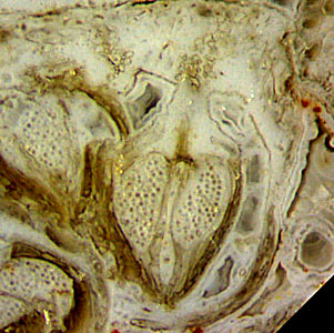
Figs.5-7: Synangia with big pedicels on cut faces of
the same chert sample as in Figs.3,4. Image sizes 1.5mm.
(Sample Bu8/18, coll. U. Wagner )
Note that the stalk may be not well seen
or not seen at all since the synangium axis most likely does not lie in
the arbitrarily chosen cut plane. More synangia with big
stalks had been found earlier in a few more chert samples
from
this location.
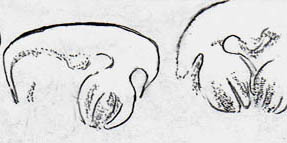
Fig.8 (below left): Pinnule cross-section drawn after the lectotype of Scolecopteris elegans
in [4], width 2mm.
Fig.9 (below right): Pinnule
cross-sections with big synangium stalks drawn after [4],
Table 5, coll. Etzold
about 1890.
Despite of these obvious
differences and other features indicating the presence of other species, M.
Barthel had stated in
[2], p.75, that most probably there is only
one maggot fern species at the type locality of Sc. elegans.
Since
the synangia in Figs.5-7 are rather similar to those in Fig.9 but
differ much from the lectotype in Fig.8, most
likely they represent a
different species. If it were one of the numerous known species
of Scolecopteris,
it should fit into one of the four groups proposed by Millay [5]
but it does not seem to fit well into any. The apically thick
sporangium wall would favour putting it together with Sc. elegans into
the "Minor group" but this is precluded by the big pedicel shaped as a
"broad cap of parenchyma uniting the sporangia to each other and to the
pinnule" [5,6]. This describes the pedicel of Sturiella
there but also fits surprisingly well to what is seen here in Figs.5-7.
Since M.
Barthel
did not notice the synangia with big stalks on the cut faces
while inspecting the sample, it
is all Sc. elegans
to him. He provides an extended description, allegedly of Sc. elegans,
based on three samples including Bu8/18, which probably
contains Sturiella only.
Such description, which probably is based on an error, is probably not
valid.
Evidence
from Figs.5-7 indicates that Figs.6-8 in [2]
and Figs.210A-G in [3] are not "certainly Sc. elegans"
but possibly Sturiella intermedia.
This would be compatible with earlier own finds of synangia with big
pedicels and of sporangia with hairs at the type locality of Sc. elegans.
In connection with
Sturiella
one need not avoid the familiar name "maggot fern" since Sturiella intermedia
(Millay
1997) is also known as Scolecopteris
intermedia
(Lesnikowska,
Galtier 1991).
It may be mentioned here that
[2] and [3] are fraught with numerous inaccurate or erroneous
size data and other irritating errors.
Example: Sc.
elegans
spore sizes in [3] are allegedly 40µm on p.228,
70µm
in Fig.209, but really 27µm. See Google:
errors palaeobotany.
There are contradictory size data on Sturiella in [5,6],
which are not relevant here.
Samples: Found by Ulrich
Wagner (Dresden) in
1997 at the type locality of Sc.
elegans and kept in his collection under the labels Bu8/23
(here Figs.1,2) and
Bu8/18 (here Figs.3-7), lent for renewed description in 2019.
H.-J. Weiss
2019
emended version
[1] E.
Zenker:
Scolecopteris elegans,
ein neues fossiles Farrngewächs mit
Fructification. Linnaea 11(1837), 509-12.
[2] M.
Barthel:
The maggot stones from Windberg ridge.
in: U. Dernbach,
W.D. Tidwell: Secrets of
Petrified Plants, D'ORO Publ., 2002. p. 65-77.
[3] M.
Barthel: Die Rotliegend-Flora
der Döhlen-Formation. Geologica Saxonica 61(2), 2015, 108-229.
[4] M. Barthel,
W. Reichel, H.-J. Weiss: "Madensteine" in Sachsen.
Abhandl. Staatl. Mus. Mineral. Geol. Dresden 41(1995),
117-135, Table 1.
[5] M.A. Millay:
A review of
permineralized Euramerican Carboniferous tree ferns. Rev. Pal. Pal.
95(1997), 191-209.
[6] A. Lesnikowska,
J.
Galtier: A reconsideration of four genera of
permineralized Marattiales ... Rev.
Pal. Pal. 67(1991), 141-152.
[7] M.
Barthel: Pecopteris-Arten
... aus Typuslokalitäten in der DDR. Schriftenr. geol. Wiss. Berlin
16(1980), 275-304.
|
 |
 24 24 |