Wilting nematophytes in Rhynie chert
(largely revised version 2020)
Nematophytes,
literally "filamentous plants", are still so poorly
understood that they go under the heading Enigmatic Organisms in [1].
Therefore, any newly observed detail is potentially relevant and worth
being reported.
There are not many details seen in the small nematophytes usually
preserved as coaly compressions [2]. 3D-preservation in chert provides
the possibility to see more. Several insights have already been
obtained from the very few nematophyte specimens found in the Rhynie
chert
hitherto. Own contributions are listed here.
The filaments of the nematophytes appear mostly as structureless tubes,
as in
Fig.1. One may ask whether it is really tubes what is seen here or
merely the tube-shaped cavities left after the organism had decayed and
vanished, together with its tube, after silicification of the space
between them. The aspect of the deformed tubes suggests that real tubes
surrounded by organic gel became silicified here.
Here the colloquial
term "wilting" is to refer to degradation and
decay of the live filaments and the organic gel in between.
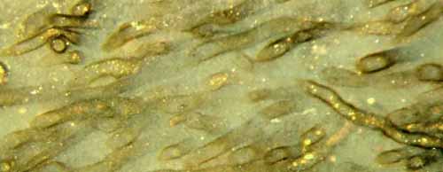
Fig.1: Nematophyte tubes in organic gel,
non-described species.
A kinked tube (above left) allows conclusions to be drawn concerning
mechanical properties. Image width 1.75mm.
Independent
of whether or not there is wall substance left in Fig.1, one can derive
information on the mechanical properties of the components before
silicification. One tube (above left) got a kink when it was bent by
contact with others. Hence, it must have consisted of a material with
some flexibility and strength. If it had been more brittle, or soft and
weak, it would have broken or torn. The gel must have been so weak that
it did not preclude kinking.
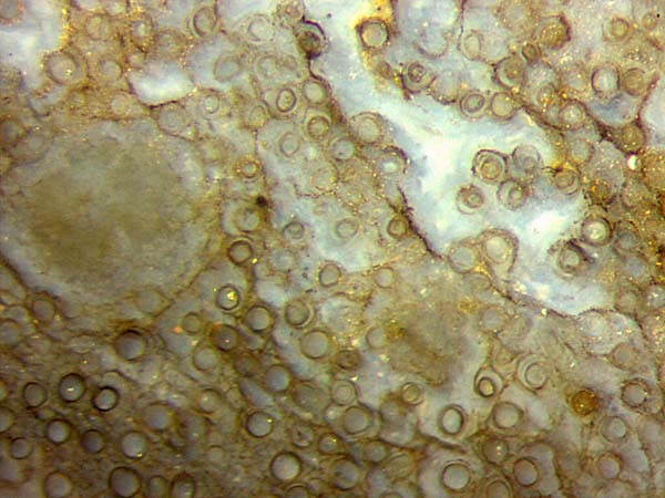
Fig.2:
Nematophyte, non-described species with uncommonly large tubes up
to 70µm across and well aligned in organic gel,
smaller ones inside indicating shrinkage. Image
width 1.4mm.
The smaller cross-sections seen inside some of the larger ones in
Figs.2 pose a problem. They could possibly be degraded and shrunken
tubes within the cavities in the silica gel preserving the size of the
original tubes. (Such phenomena are known from fossil plants, see Rhynie
Chert News 31.)
A different possibility has to be considered: The smaller section
inside may
not be the shrunken tube but the cell plasma with enclosing membrane
shrunk
away from the tube wall.
The question remains why the phenomenon is restricted to some of the
tubes in some part of the specimen. For reasons unknown,
silicification might have been not equally fast
throughout the sample so that various stages in the succession of decay
have become preserved.
Also
in Fig.2, the organic gel keeping the tubes in a compact lump below
left is seen to be degraded and fragmented above right. The decay
of gel may release some of the tubes into the surrounding water where
they float away, which explains why separate tubes are occasionally
seen scattered in the chert.
Also worth mentioning in Fig.2 are sections of two
big clots known from other nematophytes as "medullary spots", which are
assumed to somehow produce the tubes.
Fig.3 (below): Nematophyte,
same sample as Fig.1. Below
right: tubes in organic gel silicified. Above
left: gel disappeared before silicification
after an enigmatic de-jellifying front had passed, leaving
loose broken tube fills behind.
Image width 2.8mm.
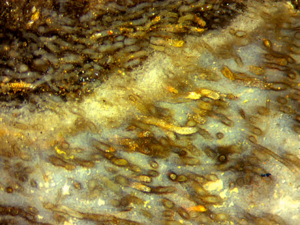
A different mode of decay is suggested by Fig.3: Apparently
the organic gel keeping the
nematophyte tube assemblage together and protecting it against
exsiccation had been dissolved by an enigmatic
decay process spreading in a front-like way, down right in Fig.3. Above left there are the
broken fills of the tubes which had settled in
the water. These
fragments indicate that there had been a transient stage of
fossilisation with silicified tube content and non-silicified organic
gel between the tubes.
Also the final stage of
fossilisation as seen in Fig.3 shows that there
had been differential
paths of silicification inside and outside the tubes, also among the
tubes, as apparent from the different colouring.
Finally it can be
stated that, before silicification, the organic gel between the
nematophyte tubes may become fragmented (as in Fig.2) or completely
removed (as in Fig.3). In either case, the tubes or fills get deranged.
Apart from these details, the nematophytes shown here are remarkable
for their comparatively huge tube diameters up to 70µm.
Samples:
Rh13/7, 0.25kg, found by S. Weiss
in 2005, Part1: Figs.1,3.
Rh2/81, 0.63kg, obtained from
Shanks in
2003, Part3: Fig.2.
H.-J. Weiss 2011, 2016,
2020
[1]
T.N. Taylor, E.L. Taylor, M. Krings:
Paleobotany,
Elsevier 2009.
[2] P. K. Strother:
Clarification of the genus Nematothallus,
J. Paleont. 67 (1993),
1090-94.
 |
 |
40 |