Ventarura
with less-common features
The Lower Devonian vascular plants preserved in the Rhynie chert,
mostly in various states of decay but also life-like, have
something in common: After the decay of soft tissue, several components
are usually left: xylem strand, spore capsules, the highly
durable spores, and occasionally the epidermis
with cuticle and a narrow strip of adjacent cortex. This
rule based on ample fossil evidence had to be
modified with the discovery of Ventarura
[1]. This plant has
invented a
unique structural element which can be conspicuous on
cross-sections as a concentric ring consisting of well-preserved cells,
positioned within the cortex between central strand and
epidermis, in the upper parts of the plant (Fig.1).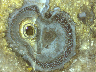
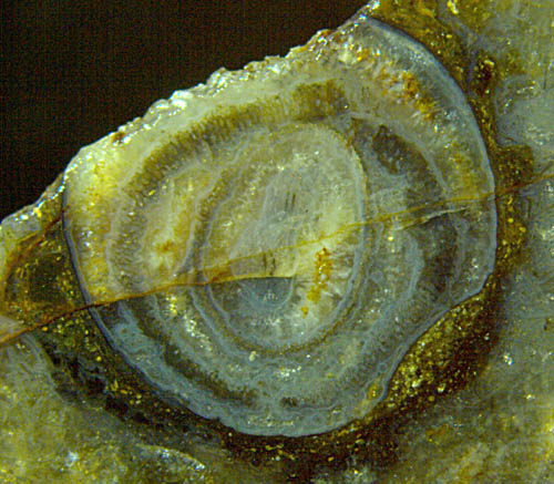
Fig.1 (right): Ventarura,
4mm across,
on a cut face of Rhynie chert, with shrivelled surface, dark ring of
well-preserved cells, xylem strand, and quartz-lined
cavity replacing part of the decayed soft tissue. Picture taken under
oil with a bubble creeping out of the cavity.
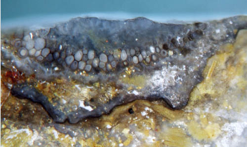
Fig.2 (left): Ventarura
fragment with distinctly seen cells of the characteristic tube.
The
well-preserved cells
forming the cylindrical tube do not always look dark and thick-walled
like
sclerenchyma. In Fig.2 they are thin-walled and mostly pale
inside.
Fig.3
(right): Ventarura,
hollow before silicification, shrivelled outside, no effect on the
characteristic cylindrical tube 5mm across,
uncommonly pale, one third broken away.
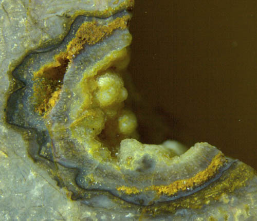 Apparently the pale tubes
are found in a few specimens only
(Figs.3-5), among them those which are not shrivelled (Figs.4,5). It is
suggested by the latter and other observations that all tubes were pale
at first and became dark later.
Apparently the pale tubes
are found in a few specimens only
(Figs.3-5), among them those which are not shrivelled (Figs.4,5). It is
suggested by the latter and other observations that all tubes were pale
at first and became dark later.
From Figs.2,3 it is also apparent that the characteristic
cylindrical tube retains its shape while the tissue within and without
vanishes. The space left by the decayed tissue between tube
and
epidermis is either left open, filled by mineral precipitate, or
vanishes by shrivelling of the decaying epidermis with cuticle.
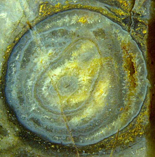
Fig.4
(left): Ventarura
cross-section of uncommon structure and preservation: 5.5mm
across, not shrivelled, two pale concentric tubes
consisting of preserved cells, the inner
one non-circular with two conducting strands inside,
quartz-lined
cavity replacing part of decayed outer tissue, yellowish quartz in
cavities
left
by decayed inner tissue.
The arrangement of tubes, on cross-sections seen as
concentric rings in Figs.4,5, the
inner one non-circular and thin, is
incompatible
with the simple forking as illustrated in Rhynie
Chert News 3.
Possibly Ventarura
is able, similar as Trichopherophyton,
to grow new sprouts within old parts: Rhynie
Chert News 82 .
The persistent tube cross-sections may not be closed rings everywhere,
as seen below in Fig.5. Note the
discontinuity of the outer tube and the combination of tube
sections with pale and darker aspect.
Fig.5
(right): Ventarura
cross-section, same shoot as Fig.4 but cut at lower
position.
Some of the facts seen in these pictures are
summarized here:
- Before silicification, the upper parts of Ventarura
had
been either shrivelled (Figs.1-3) or smooth and cylindrical (Figs.4,5).
-
The epidermis, like most of cortex, is usually not preserved but
the cuticle is seen as a dark line,
possibly a thin deposit stained
black (Figs.2-5).
- The
characteristic tube consisting of decay-resistant tissue, usually seen
as a dark ring on cross-sections (Fig.1),
can be pale and
inconspicuous.
- In case of shrivelling surface, the persistent
tube keeps its shape (Fig.3), thus appearing as mechanically
strong,
(The deviation from circular shape in
Fig.1 is
due to
reasons unknown.)
- The seeming strength of the tube is an illusion due to
the weakness of the decaying tissue around.
- There are exceptional cases of two concentric tubes,
the inner one being thin-walled.
- Irregular-shaped thick coatings on the outside
(Figs.3-5) had possibly been silica gel in watery
surroundings with abundant
debris.
Since the persistent tube tissue
is neither connected to the epidermis nor to the central strand, the
question arises how it is brought about, and for which purpose. Hence,
any finds of Ventarura
in the Rhynie chert deserve particular attention as they may lead to an
answer. Also it would be interesting to know
whether a tubular component of
the type found in Ventarura
is unique or present in some other fossil or extant plant, too.
It
is thinkable that a tube-shaped part of the cortex tissue had
been made poisonous to fend off any intruder, be it fungus or
sap-sucking creature. As a secondary effect, the tube might have become rot-resistent. Apparently the thin
persisting cell
walls had been colonized with microbial layers, thus superficially
looking
like thick walls. The seemingly thick cell walls mislead to
the assumption that the rot-resistent tube is made of
sclerenchyma [1].
A
different type of tube, a hollow straw with or without epidermis
preserved, is
often seen with Aglaophyton.
Samples: Rh19/1 (60g) Part 2: Fig.1; Rh12/35
(0.15kg) Part 2: Fig.2; both found near Smithston in 2005;
Rh4/66 (0.16kg) Part 2: Fig..3,4; Part 3:
Fig.5;
found near Smithston in 2009;
H.-J.
Weiss
2013,
modified in 2014, 2019, 2020
[1] C.L.
Powell, D.
Edwards, N.H. Trewin: A new vascular plant from the
Lower Devonian Windyfield chert, Rhynie, NE Scotland.
Trans. Roy. Soc. Edinburgh, Earth Sci.
90(2000 for 1999), 331-349.
 |
 |
58 |