Black coatings on cell walls of early
land plants
Part of the early
land plant specimens in the Rhynie chert may contain cells whose walls
had apparently become stained black (Fig.1). Closer inspection reveals
that not the walls themselves but coatings on the walls
make the black aspect, which is supported by the rare observation that
they may flake
off.
A few
selected examples are
discussed here, fitting to the more comprehensive contribution "Black
stains, coatings, and linings in the Rhynie chert", Rhynie
Chert News 83.
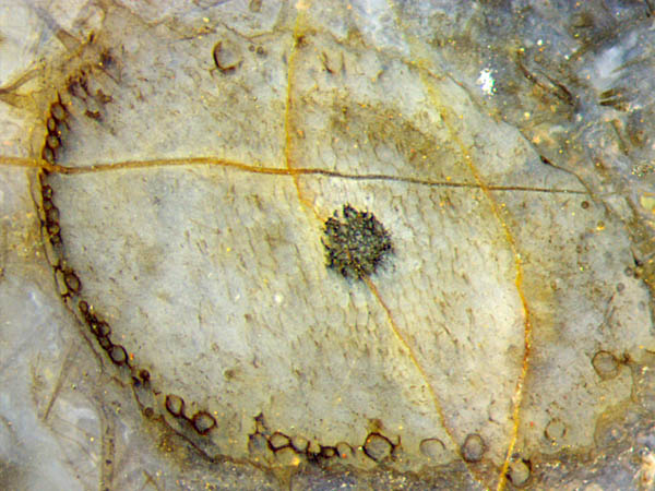
Fig.1: Trichopherophyton
section, uncommon specimen: some cell walls stained black.
Image width 2.8mm.
Trichopherophyton
usually does not show cells with stained walls, so Fig.1 shows a rare
case. It has been chosen here because it shows clearly the individual
affected cells. Mostly they are coated as a whole but a few cells below
in the picture are only partially or
weakly coated.
Much more conspicuous than the rare phenomenon in Fig.1
are the "hollow straws" [1] of the most abundant plant in the Rhynie
chert, Aglaophyton (Fig.2).
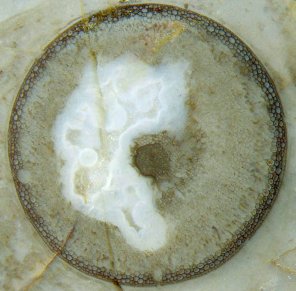
Fig.2: Aglaophyton
"hollow straw":
Peripheral layer of well-preserved cortex cells mostly with black
walls, contrasting to the decayed larger part of cortex tissue. Width
of the cross-section 4mm.
The seemingly evident explanation of the hollow
straw aspect as a result of dissolved silica penetrating into
a limited depth below the epidermis, preserving
a layer of peripheral
tissue there while the tissue farther below is left to decay
[1,2], has been doubted in [3] and is
rejected here by referring to Fig.1. There, the loosely arranged
black-walled
cortex cells are
not compatible with the idea of a silicification front.
The situation seems to be more complex, as
suggested by the observation that the well-preserved cell walls
are not always stained black (Fig.2). Apparently the
preservation as "hollow straws" is not governed by silica diffusion in
the dead plant but by the live plant providing some
kind of decay resistance to a peripheral part of the cortex
tissue. With this interpretation the question arises to which purpose
the plant had made a decay-resistant tube, seen as a dark
ring on cross-sections.
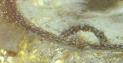
Fig.3: Aglaophyton"hollow
straw": Hole in the decay-resistant
peripheral layer repaired with a dome-shaped cap of 2mm width made from
cortex tissue.
By lucky incidence, a rare structure is documented in Fig.3: A hole in
the dark
peripheral ring of persistent tissue is seen repaired with a cap
covering the hole.
This must have occurred
while all cortex tissue was still there, because the
cap was made from cortex cells. Hence, the
ability of the plant to make part of the tissue
decay-resistant must have existed over some
span of time.
The
repair of the damage to the persistent tissue indicates that this
tissue, though only a peripheral tube a few cells thick, is of some
importane to the plant.
One may guess that not
only the black stain is a secondary phenomenon but the decay
resistance, too. Possibly
the main thing is hardly
seen here behind the secondary phenomena: The main thing might
be a repellent substance against pests applied
by the plant to a narrow peripheral region of
the cortex tissue. As a side effect, that hypothetical repellant might
preserve the cell walls, thus providing
substrates for microbes to make coatings with dark aspect.

Fig.4: Aglaophyton
sections
of quite different aspect near each other: "hollow straw"
(left) and "normal" shoot with very faintly seen tissue (right).
It has not been explained here why Aglaophyton
sometimes
appears as a hollow straw and sometimes with original tissue (Fig.4).
The phenomenon of black coatings on cell walls is widespread in the
Rhynie chert. In addition to the "hollow
straws" discussed here, black
coatings make conspicuous sights when on the cell walls of
the persistent tube of Ventarura
amidst the decayed cortex tissue.
Samples:
Rh14/18.5, obtained from Barron in 2007: Fig.1; Rh6/38.1, found in 2003: Fig.2; Rh12/162.2, found in 2007: Fig.3; Rh12/91.5, found in 2006: Fig.4;
H.-J.
Weiss
2021
[1]
C.L. Powell, N.H. Trewin, D. Edwards: Palaeoecology and
plant succession in a borehole through the Rhynie cherts, ...
Geological Society, London,
Special Publications 180 (2000), 4 39-457.
[2] www.abdn.ac.uk/rhynie
[3] A. Channing:
Processes and
Environments of Vascular Plant Silicification: Thesis, Chapter
6, Cardiff University, 2001.
 |
 |
172 |