Ventarura
with enigmatic tube inside
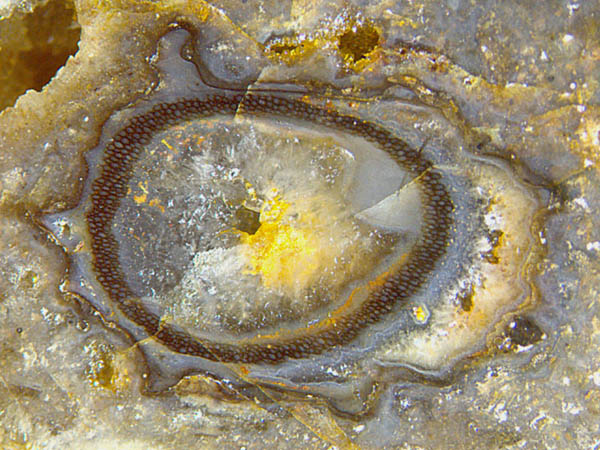 Conspicuous dark rings,
miraculously well preserved amidst largely decayed tissue
are occasionally seen on cross-sections of the early land plant Ventarura
(Fig.1). All the time while the tissue inside
the ring (or tube in 3-D) decayed and vanished and the tissue outside
the ring decayed and shrunk or vanished so that the
originally circular outer contour crumpled, the
ring itself remained unaffected, which had led to
its questionable interpretation as consisting of sclerenchymatic
tissue [1].
Conspicuous dark rings,
miraculously well preserved amidst largely decayed tissue
are occasionally seen on cross-sections of the early land plant Ventarura
(Fig.1). All the time while the tissue inside
the ring (or tube in 3-D) decayed and vanished and the tissue outside
the ring decayed and shrunk or vanished so that the
originally circular outer contour crumpled, the
ring itself remained unaffected, which had led to
its questionable interpretation as consisting of sclerenchymatic
tissue [1].
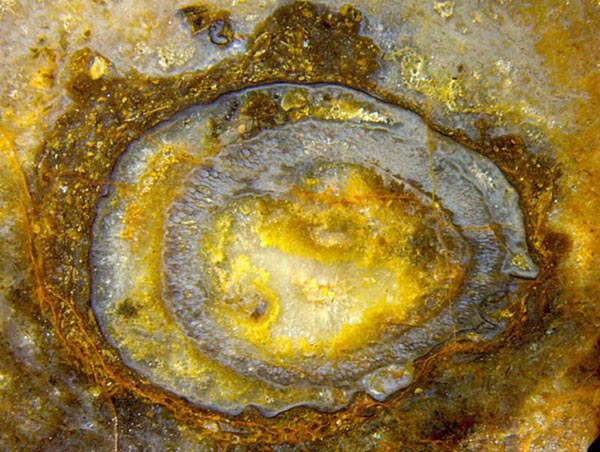
However, the rare phenomenon of apparently
thick dark cell walls locally disintegrating into dark flakes and
thin walls reveals that, despite of its
persistence and solid aspect, the tissue of the ring is not
sclerenchymatic. Also it appears that the
persistence of the ring is not coupled to some dark and solid aspect,
as seen in Fig.2. Also it
may be concluded from Figs.1,2 that the ring had essentially kept its
shape while the tissue outside became deformed or torn off.
Figs.1,2:
Ventarura
cross-sections, largely degraded and deformed before
silicification except for a tube of well preserved
tissue. Image width 10mm.
 The
persistent rings (or tubes in 3-D) pose a veritable problem. Their
persistence among
decaying tissue would
not make sense unless related to some purpose in the
live plant:
Rhynie
Chert News 58. The absence of sclerenchyma
cells implies that the tubes were not meant to stiffen the
plant. In
view of
the various inventions by early land plants to deter
herbivores, a coaxial tube of prepared tissue
might have
served the same purpose. A tube consisting of
poisonous tissue could prevent creatures from getting at the innermost
sap
ducts.
The
persistent rings (or tubes in 3-D) pose a veritable problem. Their
persistence among
decaying tissue would
not make sense unless related to some purpose in the
live plant:
Rhynie
Chert News 58. The absence of sclerenchyma
cells implies that the tubes were not meant to stiffen the
plant. In
view of
the various inventions by early land plants to deter
herbivores, a coaxial tube of prepared tissue
might have
served the same purpose. A tube consisting of
poisonous tissue could prevent creatures from getting at the innermost
sap
ducts.
As a secondary effect, the poison could possibly
prevent
decay so that the tissue persisted. As another side-effect, the
persistent
tissue offered a substrate for
microbes to form dark coatings on cell walls as in Fig.1.
The problem remains how the plant managed to prepare a definite part of
the
cortex
tissue shaped like a cylindrical tube with a substance providing
persistence.
Samples:
Rh15/28 (0.038kg), obtained from Barron in
2009, Part1: Fig.1.
Incidentally, Part2 of this sample has
provided the first
epidermis of Ventarura
ever seen: Rhynie
Chert News 61 .
Rh4/66 (0.16kg), found in 2009,
Part3: Fig.2.
H.-J.
Weiss
2021
[1] C.L. Powell,
D. Edwards, N.H. Trewin:
A new
vascular plant from the Lower Devonian Windyfield chert, Rhynie,
Trans. Roy. Soc.
Edingurgh, Earth Sci. 90 (2000 for 1999), 331-349.
 |
 |
174 |