 Trichopherophyton
aspects
Trichopherophyton
aspects
Considering the conspicuous feature of the less common but not quite
rare "hair-bearing plant", the
slender pointed bristles (Figs.1,2) whose length can exceed 1.5mm, one wonders why this plant had not been
discovered
earlier in the Rhynie chert [1]. There are possible reasons:
-
Apparently the bristles were borne as a protection against spore eaters only on the upper parts of
the plant which became less often silicified than the submerged parts.
Hence the latter (Figs.3,4) are less
easily recognized as Trichopherophyton.
- Trichopherophyton
seems to be prone to quick decay, except for the xylem strands and
sporangia, which are
not easily recognized when found scattered in the chert.
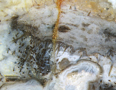
Fig.1: Trichopherophyton,
literally "hair-bearing plant", shoot
tip silicified while lying in water. (See big swamp gas
bubble below and small level bands indicating horizontal direction below
left.) The shoot seems to dive into the picture plane
towards the left, winding such that a bunch of bristles at the
very tip are directed out of the
picture plane towards the observer and thus are
seen in cross-section. Width of the picture about 7mm.
Fig.3 (below): Numerous Trichopherophyton
cross-sections
without bristles, width of
the picture 18mm.
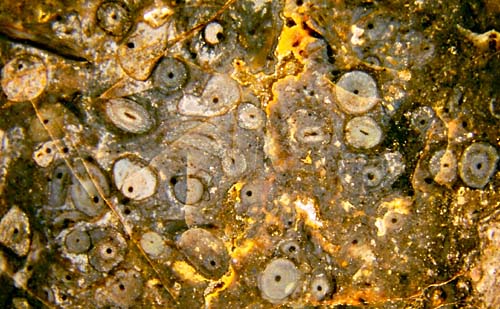
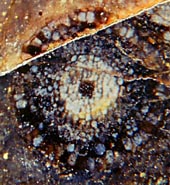
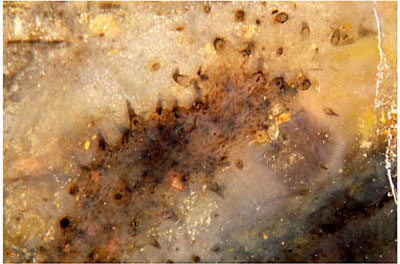 Fig.2 (right): Rare view from inside a Trichopherophyton
shoot into the bristles which are seen here as hollow cones with a
dark
dot of shrunken cell content.
Fig.2 (right): Rare view from inside a Trichopherophyton
shoot into the bristles which are seen here as hollow cones with a
dark
dot of shrunken cell content.
Fig.4 (middle): Trichopherophyton
cross-section,
width 1.5mm,
with uncommonly distinct aspect
of the different tissues. 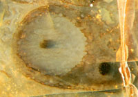
Fig.5 (right): Trichopherophyton
cross-section,
width 1mm,
grown inside an older one.
The question arises how to recognize
the "hair bearing plant" when there are no bristly shoots. Depending on
the
state of preservation, the problem is more or less difficult. A rough
rule may apply here. If any three of the features listed below are
observed, it is very probably
Trichopherophyton:
(1) circular sections, mostly 1-2(-2.5)mm
across, without occasional warts on the surface,
(2) persistent xylem, seen as
dark strands (Figs.3-6) or as conspicuous sections as in Fig.6,
(3) scalariform pattern on the tracheid walls (Fig.7),
(4) light-coloured concentric region as in Figs.4,8,
(5) new shoots developing inside older ones
(Fig.5, close-up here),
(6) detached bristles somewhere in the sample,
(7) sporangia sections of various shape, never circular, (see
Rhynie
Chert News 22).
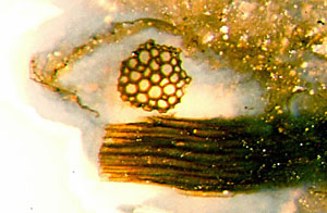
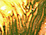 Fig.6 (far left): Trichopherophyton xylem
fragments, cross-section 0.16mm.
Fig.6 (far left): Trichopherophyton xylem
fragments, cross-section 0.16mm.
Fig.7: Scalariform pattern faintly seen in the tracheids of
the xylem strand. The divergence of the tracheids is due to the forking
of the strand.
Fig.8 (below right): Trichopherophyton
cut
lengthwise with light-coloured tissue (phloem ?) around the dark xylem
strand of 0.3mm
width. (Note also the small bluish level fill on the
left.) 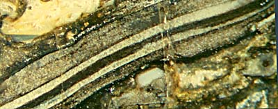
If one or more of the above features are met, one should first
make sure
that there are no features suggesting the presence of other species. (For example, (1)
combined with a wart-like outgrowth on one of the circular sections
would mean it is Rhynia.
Diameters exceeding 3mm would rule out Trichopherophyton,
etc.) Then one should
carefully inspect every inch of the sample for hidden sections with
bristles attached. In case of (5), one can be quite sure that it is
either Trichopherophyton
or Ventarura, another zosterophyll.
In the absence of fragments of the upper part of Ventarura with the
characteristic persistent tube (previously thought to be sclerenchyma) it may be difficult to tell the two
species apart.
In
view of the fact that all pictures have been taken with incident light on arbitrarily chosen cut faces,
what is seen in Fig.6 is an extremely improbable configuration: By
queer coincidence, a xylem fragment had been cut exactly across in such
a way that only a short section of a few dozen micrometers extends into depth. Against
the scattered light from the surrounding translucent
chalcedony, it looks like a piece of lace illuminated from behind. The
dark xylem fragment seen below is positioned such that it had been cut
nearly lengthwise. Considering that the fragments do not belong to any
one of the common plants in the chert but to Trichopherophyton, which had first been described as late as 1991, this image is really awe-inspiring.
H.-J. Weiss
2012, emended 2013, 2015
[1] A.G.
Lyon,
D. Edwards: The first zosterophyll from the
Lower Devonian Rhynie Chert,
Trans. Roy. Soc.
Edinburgh, Earth Sciences, 82(1991), 323-332.
 |
 |
49 |