Devonian charophyte alga forming lumps
 Charophyte algae
are well-known components of the watery habitat preserved in the Lower
Devonian Rhynie chert. One species growing whorls of branches, thus
resembling the extant charophyte Nitella,
is known as Palaeonitella
cranii [1], see Rhynie
Chert News 10,
74.
An un-named form, eventually branching at its base only, irregularly
and
without whorls, has been described in Rhynie
Chert News 48.
Unexpected recent discoveries of assumed charophyte specimes with
a whorl of branches at the top and possibly more whorls below have
been reported in Rhynie
Chert News 73, 89, 90,
93,
106,
129,
138,
139.
They have been recognized as a "Peculiar Alga" by
means of their ellipsoidal oogonia on stalks, an ancient trait
which sets them apart from all stonewort-like charophytes as we know
them.
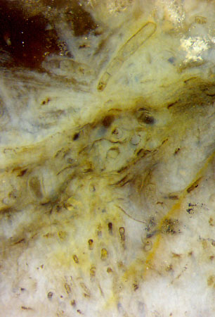
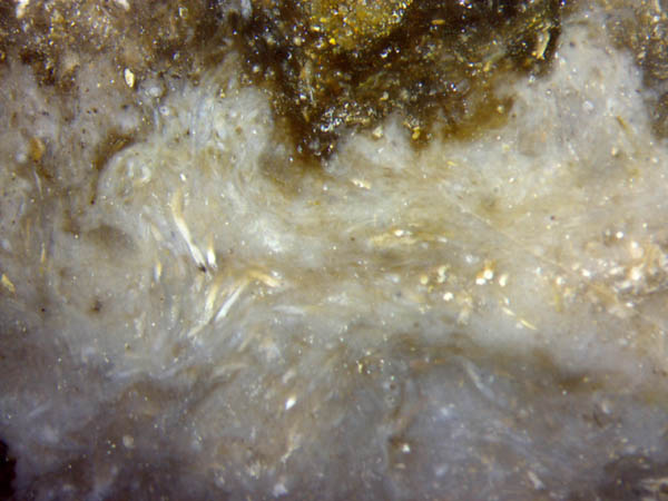
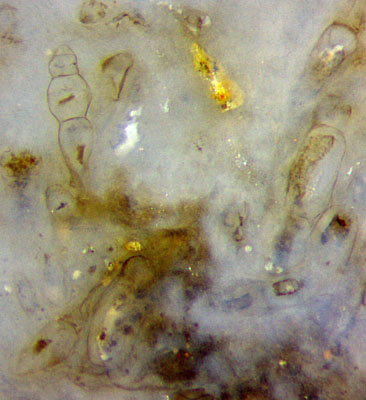
A charophyte alga growing in dense lumps of tubes (Figs.1,2),
apparently without whorls of branches, has been found in this sample,
and less clearly in a few others. Fig.1 suggests a dense
arrangement of tubes, not random but more or less correlated over some
distance, as right of the middle, where mainly cross-sections are seen.
All images from Sample Rh2/175, obtained from Shanks in 2012.
Fig.1:
Bluish chalzedony cloud in Rhynie chert, with densely grown charophyte
tubes, some with white fill; no whorls of branches. Image
height 5.2mm.
Fig.2 (far right): Aligned charophyte tubes below,
two separate tubes of 4 cells above, see also Fig.3.
Image height 2.6mm,
magnification twice that of Fig.1.
Disregarding
the confusing structure in the middle of Fig.2, there are two areas on
this cut face where the structure is easily recognized. The tubes in an
area below are so closely packed that they mimic a coherent tissue,
with cell walls hardly
visible but lengthy objects
clearly seen inside,
possibly the shrunken content of the cell.
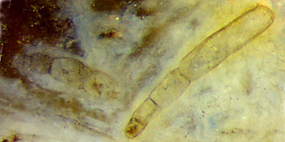
In the upper third of the
picture there are two separate tubes of
definite length,
seen enlarged in Fig.3. The slender tube on the right is clearly seen
to consist of four cells, apparently grown to the left. Less
clearly seen is the tube with four bulky cells on the left.
Fig.3 (left): Detail of Fig.2 (upper part), two separate alga tubes of
different
aspect, consisting of 4 cells each. Image width 1mm.
Same
scale as Figs.4-9.
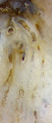
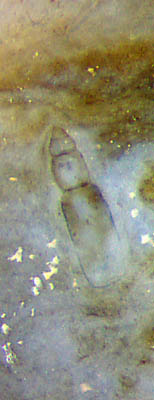
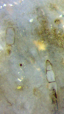
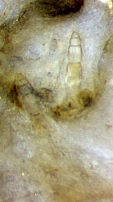
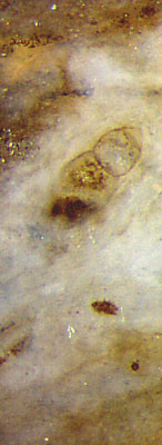
Figs.4-9:
Charophyte tube tops,
all same scale,
image height 1mm.
The diameter range of the tubes and the
articulated
structure support the assumption that the alga-like object in this
sample is a charophyte alga. It is not known
whether an alga with this particular type of growth,
with densely
packed tubes, apparently without whorls of branches, almost certainly
without gyrogonites, and with articulated tube tops as seen here, is
only a variety of the charophytes mentioned above, including
the Peculiar
Alga, or another charophyte
species. With this uncertainty kept in mind, it may be useful to
describe the facts nevertheless.
As an intriguing fact, top ends of alga tubes in this sample (Figs.3-9)
have nearly always no more than 4 cells. This agrees with the number of
cells in the branches of some charophyte whorls,
which may be incidental or not. Possibly one more cell is seen very
faintly in Fig.6 on the left tube below.
Enigmatic are Figs.3,8,
where the end wall of
the biggest cell suggests the question where it
could have broken off or how it could have formed otherwise.
As
seen in Figs.3-9, the tube tops are of variable shape, with slender or
bulky cells (Fig.3 or Figs.4,5). Very faint contours of bulky tube top
cells are also seen in Fig.9 below.
The dark matter enclosd in the
cells, diffuse as in Fig.5 or clearly shaped as in Figs.2,9, must be
the variously decayed and shrunken cell content.
The search for hidden branchings and whorls
in lumps of densely
packed tubes as in Figs.1,2 and their surroundings may contribute to
better understanding the whole.
Finally it is hoped that more details will enable this alga to be
assorted to the whorl-bearing charophyte algae
mentioned above or to a new species.
Annotation 2019: The fact that the articulated tube top parts mostly consist
of 4 cells, with the end wall of the biggest cell clearly
seen in Figs.3,8, suggests that these
parts deliberately
separated from the tubes to serve as propagules.
H.-J.
Weiss 2019
 |
 |
141 |


 Charophyte algae
are well-known components of the watery habitat preserved in the Lower
Devonian Rhynie chert. One species growing whorls of branches, thus
resembling the extant charophyte Nitella,
is known as Palaeonitella
cranii [1], see Rhynie
Chert News 10,
74.
An un-named form, eventually branching at its base only, irregularly
and
without whorls, has been described in Rhynie
Chert News 48.
Unexpected recent discoveries of assumed charophyte specimes with
a whorl of branches at the top and possibly more whorls below have
been reported in Rhynie
Chert News 73, 89, 90,
93,
106,
129,
138,
139.
They have been recognized as a "Peculiar Alga" by
means of their ellipsoidal oogonia on stalks, an ancient trait
which sets them apart from all stonewort-like charophytes as we know
them.


