Croftalania
upgrowth shaped by grazing creatures ?
As shown before, the blue-green alga Croftalania venusta [1]
can appear in surprisingly fancyful shapes as if mimicking
higher plants. Its cells adhere in a chain-like way to form filaments
which may arrange themselves into tufts resembling
bushes or trees. The tufts of filaments grown in water, as seen
in Fig.1, look quite natural and do not give rise to wonder
but
they do when shaped like those in Figs.2,3 (same scale). Really,
pointed
shapes with smooth boundary are seen more often than the ones without
such attributes requiring an explanation.
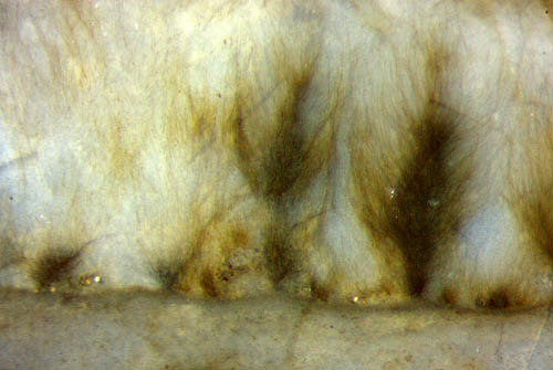
Fig.1: Blue-green alga
Croftalania,
filaments grown
in water arranged in tufts resembling
bushes or trees. Image width 2mm.
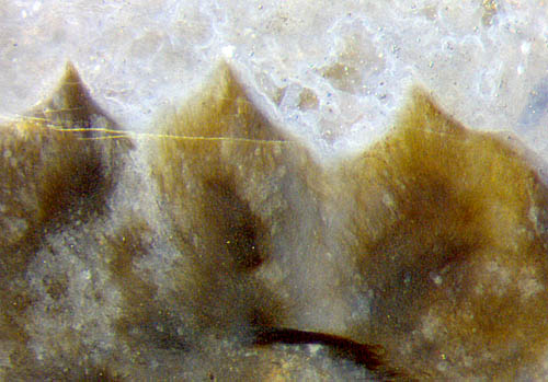
Figs.2,3 (below Fig.1): Croftalania tufts with distinct
contour. Image width 2mm.
 A moult part of the small crustacean Castracollis
seen
close to trimmed Croftalania
has led to the proposal that the creature could
have nibbled away the loose ends of the filaments, thus making a smooth
contour (Rhynie Chert
News 56).
This seems to be
confirmed by another sample of Rhynie chert, presented
here, with a similar constellation of such parts
(Fig.4). Here, a large moult part from a rather
big specimen of Castracollis,
with the typical segmentation, 14 segments visible, is found
among large amounts of
shaped Croftalania
, part of which is seen in Fig.4 (in the upper left corner) and in
Fig.3, where Croftalania
invokes
the illusion of a mountain range. The smooth contours in Figs.2-4
suggest the presence of a binding organic gel between the filaments,
keeping the whole in shape.
A moult part of the small crustacean Castracollis
seen
close to trimmed Croftalania
has led to the proposal that the creature could
have nibbled away the loose ends of the filaments, thus making a smooth
contour (Rhynie Chert
News 56).
This seems to be
confirmed by another sample of Rhynie chert, presented
here, with a similar constellation of such parts
(Fig.4). Here, a large moult part from a rather
big specimen of Castracollis,
with the typical segmentation, 14 segments visible, is found
among large amounts of
shaped Croftalania
, part of which is seen in Fig.4 (in the upper left corner) and in
Fig.3, where Croftalania
invokes
the illusion of a mountain range. The smooth contours in Figs.2-4
suggest the presence of a binding organic gel between the filaments,
keeping the whole in shape.
The gray spots partially obstructing the sight of the filaments in
Figs.2,3 seem to be due to secondary phenomena related to decay
A few specimens of another small crustacean, Ebullitiocaris,
which is smaller than Castracollis,
have
been found in the same chert sample (Fig.5).
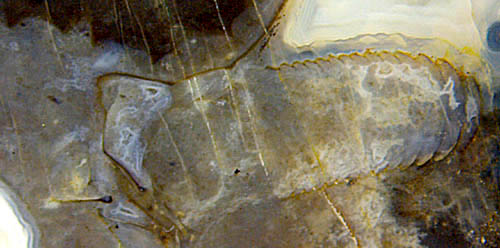
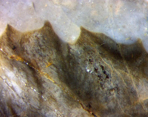
Fig.4 (left):
Crustacean Castracollis
moult part with
typical segmentation; Croftalania
(dark) with pointed contour nearby.
Image width 6mm.
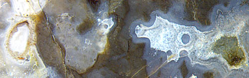
Fig.5 (left):
Crustacean Ebullitiocaris
(far left), segmentation hidden inside, not seen;
Croftalania
(dark) nearby.
Same scale as Fig.4.
The
fossil evidence leads to the conclusion that the smooth contour with
cusps had most probably been brought about by crustaceans
grazing on Croftalania
filaments embedded in organic gel.
Samples: Fig.1: Same sample as in Rhynie Chert
News 56, own find in 2001, labelled Rh2/19.2;
Figs.2-5: Taken from a sample in the collection of Steffen Koehler, Meissen, collected decades ago by Brian Beveridge, Gloucester,
on the now protected site formerly owned by A.G. Lyon, Rhynie, documented here under the label Rh2/302.1 .
H.-J.
Weiss
2018
[1] M. Krings, H. Kerp, H. Hass, T.N.
Taylor, :
A filamentous
cyanobacterium showing structured colonial growth from the Early
Devonian Rhynie chert.
Rev. Palaeobot. Palyn.
146(2007), 265-276.
 |
 |
125 |