Nematophytes vs. liverworts
Nematophytes,
which literally means "filamentous plants" (Fig.1), are
still so poorly understood that lately they have been tentatively
listed under the heading "Enigmatic Organisms" [1]. This poses a
challenge to look for explanations [2].
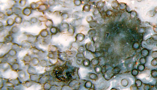
Fig.1: Nematophyte in Rhynie chert with nearly parallel tubes in
cross-section and two dark clots with obscure inner structure; undescribed species. Width of the picture 1.26mm.
(Annotation
2020: Obviously there is no tangle of tubes inside the clots, perhaps with the exception of very small ones not seen here.)
Recently it was tried, with much effort, to gather evidence for the
idea that some or most nematophytes are ancient liverworts [3]. That
idea is based on two guesses:
- The tubes constituting the nematophyte are
rhizoids of some liverwort.
- The cuticles with cellular patterns known as
Nematothallus
[4,5]
and Cosmochlaina,
often found
together with coalified tangles
of filaments or tubes, belong to ancient
liverworts.
The idea of a liverwort connection has quickly spread among the
scientific community and more or less been accepted without checking,
judging from quotations like this: "One interesting hypothesis suggests
that several of the enigmatic Cambrian to Devonian fossils
traditionally included in the nematophytes may represent remains of
ancient liverworts ... [1]." Apparently this hypothesis has never been
seriously doubted, except for the present contribution based on a very
few nematophyte specimens recently found
in the Rhynie chert [6].
The dark clots among the mass of tubes in Fig.1, which are a typical
feature of some nematophytes, preclude an interpretation of the tubes
as rhizoids.
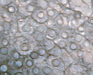
Fig.2: Same sample as Fig.1, with a cellular pattern interpreted here
as boundaries between gel coats around the tubes. See also Rhynie
Chert News 30
The problem of how the nematophytes could possibly produce a cuticle
with cellular aspect can be separated into two parts: How the pattern
is brought about, and how the cuticle is made.
Fig.2 suggests an
explanation for the origin of the pattern: Every one of the tubes
surrounds itself with a coat of gel. The gel coats taken together make
a continuous mass of gel, with the boundaries between them still there
and, under favourable conditions, seen as a cellular pattern.
It remains to be explained how the pattern can be imprinted onto a
cuticle. This is attempted with the drawing in Fig.3. Here it is
assumed that the boundaries between the gel coats seen on a cut and
polished chert face in Fig.2 make a network of grooves on the surface
of the lump of gel, possibly deepened by drying.
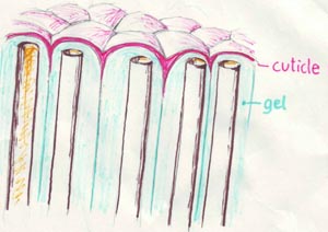
Fig.3: Schematic drawing illustrating the possible formation of a
cuticle with cellular pattern (Nematothallus
aspect) on the gel surface
of a nematophyte. (The shape of the tube ends is not known, here drawn as open for 3D-illusion.)
The tubes may produce a cuticle precursor substance and release it into
the gel where it moves by diffusion towards the surface where it
accumulates, spreads, fills the grooves, and polymerizes into a
protective cuticle with Nematothallus
aspect, which is decay-resistant
as known from the cuticles of land plants.
This is the proposed solution of the long-standing Nematothallus
problem, without invoking the idea of a liverwort connection. It is
essentially based on the presence of gel as a constituent of
nematophytes which reveals itself in the Rhynie chert but not with the
compressed specimens found elsewhere.
A similar
solution is proposed for the Cosmochlaina
problem.
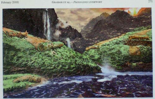
Fig.4: Prototaxites
forming from liverwort rolls as imagined by
Graham
et al. (2010) [7].
The vanishing evidence for the liverwort interpretation of nematophytes
affects the latest fancy hypothesis declaring the enigmatic huge
Prototaxites
trunks to be rolled-up and subsequently silicified liverwort mats as
illustrated in Fig.4 [7].
The unique preservation of nematophytes in the Rhynie chert,
non-compressed and suggesting the presence of gel between the
filaments, allows the following conclusions to be drawn [6]:
- The idea of nematophytes being related to
liverworts, brought up in 2004 and widely spread since, is
not substantiated.
- The latest elaboration of the liverwort idea in
2010, which is the introduction of liverwort rolls as another
interpretation of
Prototaxites,
might be remembered for its inventiveness but should
better be forgotten.
H.-J. Weiss
2010 2016 2020
[1] T.N.
Taylor, E.L. Taylor, M. Krings:
Paleobotany,
Elsevier 2009,
p163.
[2] H.-J.
Weiss : "Enigmatic Organisms" – Insights derived
from new
finds. (Poster)
8th European
Palaeobotany - Palynology Conference 2010, Budapest.
[3] L.A.
Graham, L.W. Wilcox, M.E. Cook, P.G.Gensel :
Resistant tissues of
modern marchantoid liverworts resemble enigmatic Early Paleozoic
microfossils.
Proc. Nat. Acad.
Sci., USA, 101(2004), 11025-29.
[4] P.K.
Strother : Clarification of the genus
Nematothallus.
J. Paleont. 67 (1993), 1090-94.
[5] H.
Steur, W. v.d.Brugghen : Nematothallus – een
raadselachtige plant
uit het Siluur en het
Vroeg-Devoon. Grondboor & Hamer (1998) Nr.2, 28-35.
[6] H.-J.
Weiss : Nematothallus:
How the filaments produced a cellular cuticle. (Oral presentation)
8th European
Palaeobotany - Palynology Conference 2010, Budapest.
[7] L.E.
Graham, M.E. Cook, D.R. Hanson, K.B. Pigg, J.M. Graham:
Structural, physiological, and stable
carbon isotope evidence that
the
enigmatic Paleozoic fossil Prototaxites
formed from rolled liverwort
mats.
Am. J. Bot. 97(2010), 268-275.
Acknowledgement: Thanks are due to Christopher Taylor ,
Curtin
University, Perth,
for drawing attention to the above painting and related discussions by
means of his blog, which instigated the presentation [6].
 |
 |
38 |