Palaeozoic microbes in
chert
Microbes
grown in swamp water can be found preserved in chert. The
freshwater cyanophyte Croftalania
[1] forms filaments and tufts with sizes of about 1mm: Figs.1,2.
(See also Rhynie
Chert
News 56.)
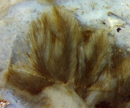
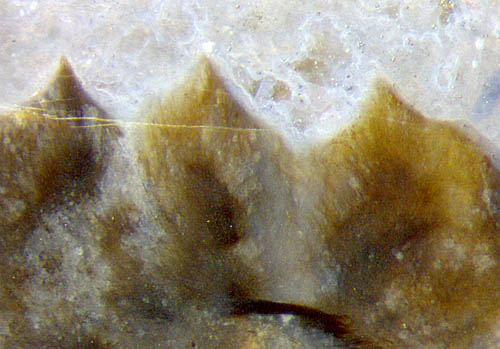 Fig.1:
Tufts of the cyanophyte
Croftalania
in the Devonian Rhynie chert. Width of the filaments about 3µm [1].
Image width 2mm.
Fig.1:
Tufts of the cyanophyte
Croftalania
in the Devonian Rhynie chert. Width of the filaments about 3µm [1].
Image width 2mm.
Fig.2: Tufts
of the cyanophyte
Croftalania:
surface apparently shaped by grazing creatures. Image
width 2mm.
The peaks in Fig.2 do not look like mere tufts
of cyanophyte filaments
in water. Their smooth surface seems to have been shaped at
a stage when the filaments were fixed in gel and grazing
creatures were still moving around in the water before all became gel
and finally chert.
Distinctly
different from the tuft-like growth of Croftalania
is the growth mode
producing stacks of thin sheets (Figs.3-6). Often
they are nearly plane or slightly curved, also broken. The
layered stacks roughly resemble wood fragments cut
lengthwise but their occasionally seen
delaminating ends and their connection to randomly shaped flakes or
lumps in the water of the swamp suggest a microbial origin
(Fig.3). (See Permian Chert
News 43.)
The crack in Fig.4
shows that the sheet
stack had been
stiffenend by early silicification, broken, then fused with the
surroundings and hardened by
continuing silicification.
Layered deposites of mineral matter mediated by
microbes are well known as stromatolites [2] formed in
shallow salty water or in freshwater
[3].
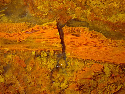
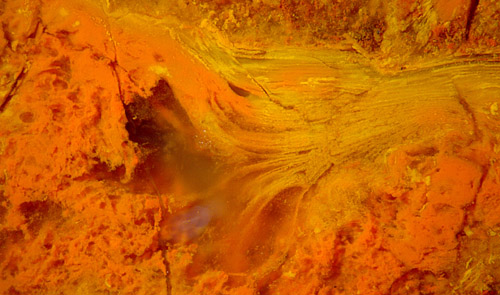
Fig.3: Stack
of microbial sheets in Permian red chert, delaminating while
soft and merging with
irregular-shaped microbial lumps on the
left. Image width 5.5mm.
Fig.4 (far right): Stack
of microbial sheets in
Permian red chert, broken
while the surrounding swamp matter was still fluid. Image
width 5.5mm.
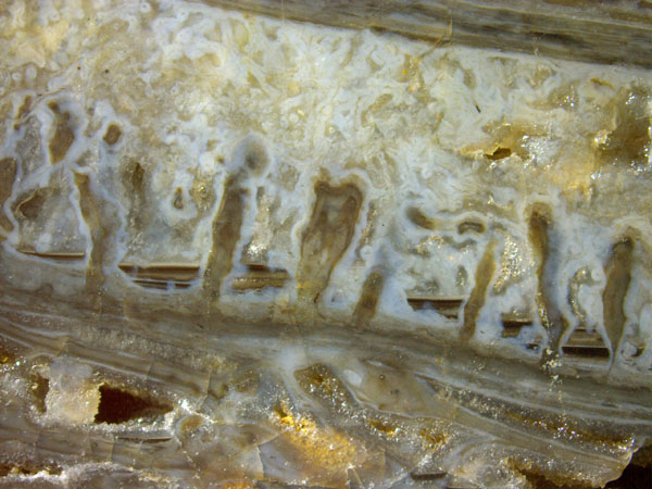
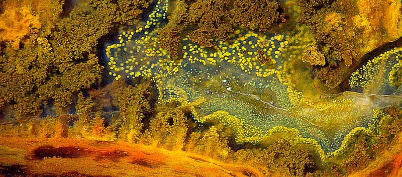
Fig.5: Microbes in Rhynie chert arranged as large flat sheets with
emergences therefrom, dark level fills from fluid
suspensions. Image width
17mm.
Emergences may arise from large sheets
of microbial origin (Fig.5). The
little black pools with horizontal surface in between are former
troughs where a heavy watery suspension involving dark microbes (?) had
accumulated and separated itself from the water above before all turned
into gel, then into chalcedony.
Fig.6 (below): Red Permian chert: laminated layer
with tunnels eaten by elusive creatures;
silica precipitates, probably induced
by microbes,
including emergences and clouds above. Image width 3.5mm.
Apparently the emergences in Fig.6 are closely
related to the
laminated red substrate below, which is a stack of microbial sheets as
in Figs.3,4.
The crack in Fig.4 runs across a stack of microbial
sheets but not farther, which indicates that the surrounding swamp
matter was still fluid. Hence,
early silicification making the stack solid must have been
caused by the microbes. Thus it
can be assumed that in other cases, too, microbes
do not only passively get trapped in silica gel but promote
silicification.
The microbes involved in
the formation of the structures shown in Figs.3-6 have not been
identified here.
.
H.-J.
Weiss 2023
[1]
M. Krings, H. Kerp, H. Hass, T.N. Taylor, N.
Dotzler:
A filamentous
cyanobacterium showing structured colonial growth
from the
Early Devonian Rhynie chert.
Rev. Palaeobot.
Palyn. 146(2007), 265-276.
[2] T.N.
Taylor, E.L. Taylor, M. Krings: Paleobotany,
Elsevier 2009, 115-117.
[3] P.
Freytet, E. Verrecchia: Freshwater organisms that build
stromatolites: A synopsis ... Sedimentology (1998), 45,
535-563.
 |
 45 45 |