How "coprolites" grow
One piece of petrified wood
challenging the
"state of the science"
Dark clots like those in Fig.1 have been found in petrified wood
worldwide and interpreted as coprolites of small creatures in numerous
publications. Remains of creatures of fitting size have never been seen
near the clots. Nevertheless the imaginary creatures have been
specified in [1] and previous publications as oribatid mites, also more
cautiously as "unknown creatures" [2] or "new detritivores" [3].
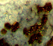
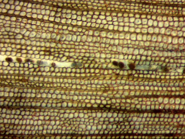 Fig.1: Silicified wood,
cross-section, one radial row of tracheids partially replaced by
dark clots.
Fig.1: Silicified wood,
cross-section, one radial row of tracheids partially replaced by
dark clots.
Lower Permian, Schallodenbach,
Rhine-Palatinate. Width of the picture 1.4mm.
Fig.1 illustrates one of several reasons for doubt concerning the
current interpretation: No creature would have been able to neatly gnaw
off a row of wood cells and replace them with same-size coprolites.
Doubts of this kind were ignored by the proponents of the coprolite
hypothesis since 2007.
For lack of reasonable arguments, R. Rößler, author and co-author of
numerous publications on the alleged coprolites, involved a lawyer to
declare the coprolite interpretation of dark clots as the current
"state of
the science". To think of this ! Those who shy away
from discussions try to define the "state of
the science"! This is contrary to the very principles of
science
and therefore cannot be kept up for long. So it is not really
surprising that it takes only one image of petrified wood (Fig.2) to
show how the clots are formed and to expose
the coprolite interpretation
as nonsense.
(The other images are for additional substantiation. All
images are of the same scale. They show the polished face of a sample
cut off from a larger find kindly provided by Ch. Krüger,
Schallodenbach. It is kept in the own collection
under Sch/3.1. The larger part has been returned.)
Fig.2 (below): Silicified wood with pale clots inside cells
seen
to be evolving into dark clots in destroyed tissue: wood rot in
progression. Width of the picture 1.4mm.
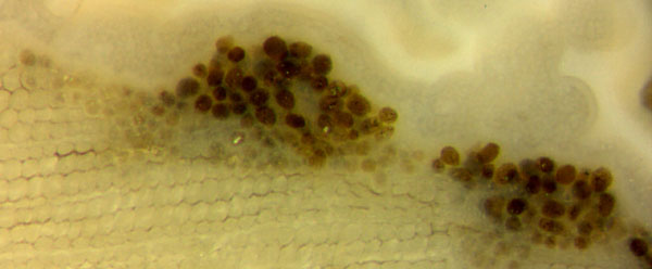 Fig.2 indicates that pale
clots originate within the apparently empty cells and grow
darker. This
reveals the succession of events: Mechanical damage of the
wood
favouring the onset of rot beginning with pale clots in the cells near
the fracture face, proceeding with the decay of cell walls and
darkening and eventual expansion of the clots. The same occurred at
several places within this sample, see Fig.3.
Fig.2 indicates that pale
clots originate within the apparently empty cells and grow
darker. This
reveals the succession of events: Mechanical damage of the
wood
favouring the onset of rot beginning with pale clots in the cells near
the fracture face, proceeding with the decay of cell walls and
darkening and eventual expansion of the clots. The same occurred at
several places within this sample, see Fig.3.
Fig.3 (below): Wood rot in progression (upwards
in this image). Width of the picture 1.4mm.
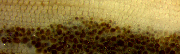 Apparently
the partial processes mentioned above are not always coupled: Pale
clots may accumulate in large numbers without turning dark (also in
this sample but not shown here), and dark clots may be found in parts
of tissue without damaged cell walls (Figs.3,4). This implies that dark
clots have not always expanded beyond cell size (Figs.3-5).
Apparently
the partial processes mentioned above are not always coupled: Pale
clots may accumulate in large numbers without turning dark (also in
this sample but not shown here), and dark clots may be found in parts
of tissue without damaged cell walls (Figs.3,4). This implies that dark
clots have not always expanded beyond cell size (Figs.3-5).
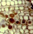
Fig.4 (far right): Dark clots in well-preserved tissue. An
interpretation as coprolites is obviously excluded for several reasons.
Width of the picture 0.25mm.
 It
be mentioned here that clots of this origin are seen not only as
globules within cells or among damaged tissue but also as angular
replicas of the cell lumina as in Fig.5., where the dark matter
apparently has filled the cell completely and kept this shape after the
decay of the cell walls. Sometimes such replicas are the only
structural information left of the vanished tissue, as in the present
case. Even angular cell-size clots have repeatedly been mistaken for
coprolites [4].
It
be mentioned here that clots of this origin are seen not only as
globules within cells or among damaged tissue but also as angular
replicas of the cell lumina as in Fig.5., where the dark matter
apparently has filled the cell completely and kept this shape after the
decay of the cell walls. Sometimes such replicas are the only
structural information left of the vanished tissue, as in the present
case. Even angular cell-size clots have repeatedly been mistaken for
coprolites [4].
Fig.5: Angular replicas of pith cells with
polygonal cross-sections: only evidence of the former tissue structure
of the decayed central pith. Width 0.4mm.
The above pictures taken from one sample which is representative of all
silicified wood with cell-size clots lead to the following conclusions:
(1) Every dark clot grew from a pale clot within a cell.
(2) The clots represent a kind of wood rot which can destroy the tissue.
(3) After decay of the cell wall the clots can expand and displace
themselves so that the wood structure gets lost.
(4) Cell-size clots in silicified wood, arranged in rows or at random,
have
lightly been interpreted as coprolites.
(5) The interpretation as coprolites in all
related publications, including [1-4], is utterly wrong.
H.-J.
Weiss
2015
[1] Z.
Feng, J.W.
Schneider, C.C. Labandeira, R. Kretzschmar, R.
Rößler:
A specialized
feeding habit of Early Permian oribatid mites.
Palaeogeography,
Palaeoclimatology, Palaeoecology 417(2015), 121-124.
[2]
M. Barthel, M. Krings, R. Rößler: Die schwarzen Psaronien
von
Manebach, ihre Epiphyten, Parasiten und Pilze. Semana 25(2010), 41-60.
[3] Zhuo
Feng,
Jun Wang, Lu-Yun Liu, R. Rößler:
A novel coniferous tree trunk with septate pith ..: -
Ecological
and evolutionary significance.
Int. J. Plant Sci.
173(2012), 835–848.
[4] R.
Rössler: The late palaeozoic
tree fern
Psaronius - an ecosystem unto itself.
Rev. Palaeobot.
Palyn. 108(2000), 55-74.
|

|
 26 26 |