Alleged borings in fossil
wood and their possible formation
Small areas of vanished tissue in coniferous fossil wood have
repeatedly been explained with the wood-eating habit of oritabid mites
[1] or unknown creatures [2], which has been refuted in Fossil
Wood News 4
and 5.
Although it is easy to explain why the small cavities are
not what they are usually thought to be, it is not obvious how they
really could have been brought about. The presence of dark clots
thought to be coprolites was offered as evidence supporting the
interpretation as frass galleries. Thereby, contrary evidence was
ignored: Often the alleged frass
gallery is not wider than the alleged
coprolite. Also the size and shape of the alleged
coprolites often fit to the size and shape of
the cells, and the clots may be found inside
cells.
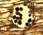
Fig.1
(right): Alleged frass gallery with coprolites in Lower Permian
coniferous wood, detail from Fig.17 in [2]. Obviously the alleged
coprolites are cells filled with dark matter.

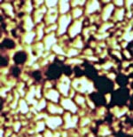
Fig.2
(left): Alleged tunnel eaten by oribatid mites into
coniferous
wood, with angular clots interpreted as coprolites in [1], detail from
Fig.3C in [1].
While Fig.1 may be explained by the presence of
an infectious agent, a kind of wood rot, spreading successively
from cell to cell, Fig.2 seems to require a more complex
explanation.
The angular shape of the clots suggests that they, too, once had been
confined to cells like those in Fig.1 and released when the cell walls
decayed. What is unusual in Fig.2 is the circular section of
the cavity, which is usually not observed in connection with cell-size
angular clots. Hence it is interesting that there is a
process of obscure nature which makes round holes into the wood, as
seen in Fig.3. 
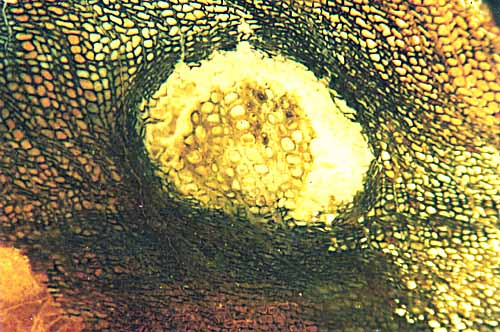
Fig.3:
Largely deformed wood by local expansion of unidentified cause. Note
the continuity of the cell files where the bloated wood is connected to the compressed wood below. Width
of the cavity 2mm.
Obviously the wood inside the cavity is bloated while the surrounding wood is compressed, with a wide enigmatic
gap which is not fully circular. Probably
the deformation did not occur in the living tree but in the dead wood
with the tissue still coherent but its strength reduced due to degradation.
The compression can be traced back to two components: the radial expansion
of
unknown cause and a superimposed shear deformation of the wider
vicinity. The shear was of such orientation as to exert pressure from
above right and below left, which manifests itself by the concentration
of compressed tissue there and possibly also by the not quite circular
section.
Also remarkable is the continuity of the cell files where the bloated
tissue is connected to the compressed tissue below.
These
observations are not sufficient to explain what had been going
on here before silicification of the whole. One can only guess
that it is another one of the various examples of fungus or microbial
action.
Anyway, here is evidence that small holes in wood are not always
borings. This supports the assumption that
most of the alleged borings and frass galleries are no such, and that
the cell-size clots seen there are no coprolites. Neither oribatid mites nor
unknown creatures need be invoked to explain certain types of damage in
Palaeozoic wood.
Sample of Fig.3: pebble found in 1994 in a dry creek in Utah (W 111°58', N 37°8' ), possibly from Chinle formation.
H.-J.
Weiss
2012
[1] Zhuo
Feng,
Jun Wang, Lu-Yun Liu :
First report of oribatid mite (arthropod) borings and coprolites in
Permian woods from the Helan Mountains of
northern China.
Palaeogeography,
Palaeoclimatology, Palaeoecology 288(2010), 54-61.
[2] M. Barthel, M.
Krings, R. Rößler: Die schwarzen Psaronien von
Manebach, ihre Epiphyten, Parasiten und Pilze,
Semana 25(2010), 41-60.
(For related references see Fossil Wood News 3-8.)
|

|
 14 14 |

 14
14


 14
14