Rhynia aspects
Rhynia gwynne-vaughani
had been known as the less abundant
and smaller one of the two early Devonian land
plants named Rhynia
until the bigger one had got a new name, Aglaophyton. Among
the mostly deformed or partially decayed sections seen in the Rhynie
chert, Rhynia is
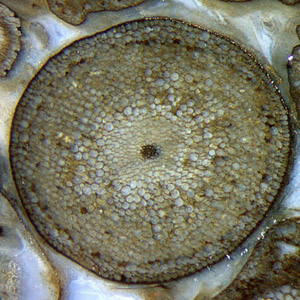
more likely to be seen with a few well-preserved specimens like the one
in Fig.1. It is an uncommonly large
one, 2.6mm across.
Most diameters are well below 2mm. The
smallest ones in Fig.2 could be Rhynia
gametophytes. Fig.1 may serve as an
introductory image here while the following ones taken from another
sample are to draw attention to various other observations.
Note
also the vaguely seen crack entering at the top of Fig.1, being
deflected at the cuticle on the plant surface, running there along for
a quarter of
the circumference, and departing from the surface on the right. The
thus debonded part of the surface is marked by a
row of tiny bright dots. The waxy
cuticle, which covers all land plants as a
protection against exsiccation, provides an easy crack path in the
silicified state.
Fig.1: Rhynia gwynne-vaughani
well preserved among deformed shoots, cross-section
2.6mm,
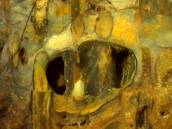
Fig.2: Surface of a Rhynie chert sample with a former bubble
in the
swamp, now seen as a cavity with thickly coated Rhynia.
Image width
17mm.
Most conspicuous on the natural surface of this
Rhynie chert sample of
0.3kg (Fig.2) is the
cavity with two thickly coated Rhynia
shoots seen inside. Incidentally they are also seen as inclined
sections
in the compact chert above. The cavity had been a bubble in the swamp
water, possibly oxygen
produced by algae or swamp
gas,
trapped among plant shoots and microbial layer stacks, some
of the
latter hidden in the bright-coloured areas above and elsewhere.
Former
bubbles now seen in chert had become stabilized by silica gel formation
around them, then filled with silica-rich water while the gas had
escaped by diffusion before silicification.
There are more than
one indications that silicification processes had been
going on
within the water-filled bubble. First came a thin dark lining, not well
seen here, possibly from a
thin microbial lining on the cavity wall. The small yellow deposit at
the bottom is probably from tiny
silica grains raining down from the water where
they had formed. (The grains could have grown suspended in gel which
liquefied later under changing parameters like temperature or pH.)
Then, possibly triggered by substances released from the
decaying Rhynia
shoots in the bubble,
thick coatings of silica gel formed around them. With
time,
the silica gel gradually turned
into chalcedony. A crack and small
displacement in the shoot on the
left
indicate fracture while the whole was not yet fully hardened.
The remaining water escaped by diffusion so that
part of the former bubble is empty space now.
What looks like bulging eyes on a downward
creeping snake above left in Fig.2 is
the typical warts often seen on Rhynia
shoots, whose purpose is not quite obvious.
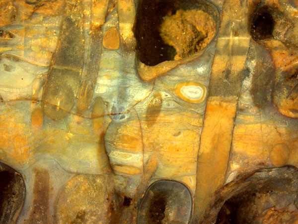 Fig.3 (right): Raw
lateral face of a Rhynie chert layer
fragment with former
bubbles stuck among Rhynia
and microbial sheets; upright Rhynia with
levels inside. Image width 17mm.
Fig.3 (right): Raw
lateral face of a Rhynie chert layer
fragment with former
bubbles stuck among Rhynia
and microbial sheets; upright Rhynia with
levels inside. Image width 17mm.
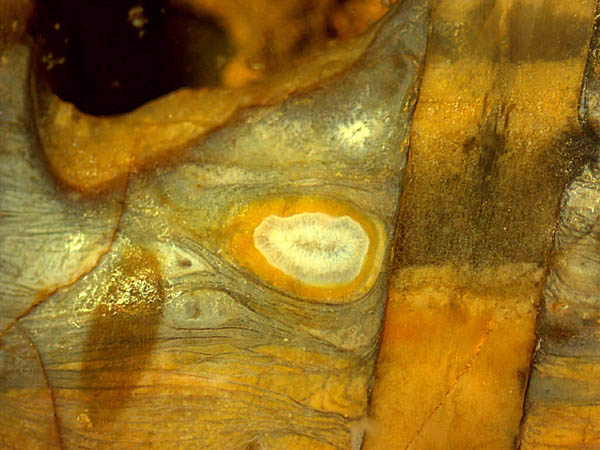
Fig.4 (below): Enlarged part of Fig.3, with
peculiar details
clearly visible: levels inside Rhynia,
agate inside bubble, microbial sheets, and others. Image width 7mm.
What may appear confusing in Fig.3 can be discussed more
easily in
Fig.4. More conspicuous but less problematic is the agate fill
of a small former bubble. One may only wonder why it is
the only
one of its kind in this chert sample.
More problematic is the sequence of silicification stages forming the
various levels inside the Rhynia
shoot. Levels of this kind, indicating the
horizotal direction during silicification, are
usually
the result of settling emulsions or suspensions in cavities. Judging
from Fig.5, decaying plant tissue did not much interfere with
the
process. From the fact that the levels are
confined to
the interior of plant shoots (or bubbles) it
can be concluded that the silicification processes inside those
compartments went on independent of what had been going
on outside. Microbial sheets formed in
the swamp water are
clearly seen in cross-section
as thin dark lines in Figs.4,5. The brown spot
on
the left in Fig.4 must be some kind of stain which had got
there later since it does not interfere with
the microbial sheets. It remains unexplained
here.
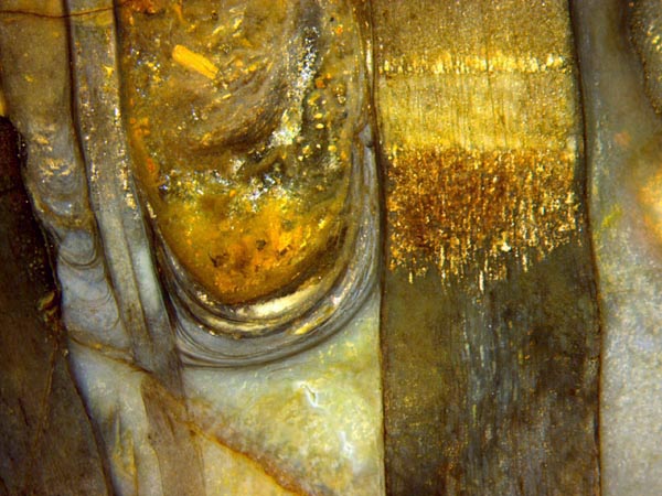
Fig.5: Cut face of the same Rhynie chert sample as above; two upright
Rhynia
shoots of remarkably differing diameters, 0.5mm and 2.2mm, with
a stack of microbial sheets suspended between them, forming a trough
later filled with muddy water apparently flooding the partially
silicified swamp. Image width 7mm.
Like
some other phenomena revealed in these pictures, the conspicuous trough
in Fig.5 does not suggest an obvious explanation. The variable contact
angle on the left precludes the simple explanation as microbial sheets
grown on a meniscus of a liquid. As a possible
but not quite consistent explanation, a
sinking water level or drying silica
gel caused the microbial
sheets grown between upright Rhynia shoots to sag,
and later all became
flooded with muddy water from elsewhere, judging from the
stack of yellow
mica platelets and other debris.
It is hard to imagine how the trough might be shaped behind the Rhynia shoots in
Fig.5 and how it could be compatible with the small trough-like sheets
on the left of the smaller Rhynia.
Instead of grinding Fig.5 away in order to see what lies behind,
samples with similar phenomena will be inspected.
Samples:
Fig.1: Fragment of a chert layer of
13cm, 0.7kg, 1998
obtained from Margaret Shanks, labelled
Rh2/5, here cut face
of Part1,
Figs.2-5: Chert sample
of 0.3kg, 2009 obtained from Barron jr.,
labelled Rh15/6, here
surface and cut face of Part1.
H.-J.
Weiss
2018
 |
 |
127 |