Nothia
sporangia aspects
Among the early land
plants found in the Rhynie chert, Nothia
is distinguished by big tubes, called giant cells in [1], embedded in
the epidermis. Since they are no mere giant cells but tubes formed by
dissolution of numerous cell walls, as
extant euphorbias do to store a poisonous milk there, it had been
proposed here that Nothia,
too, had formed the tubes as a defense
against herbivores. Usually these tubes are not well seen, as in
Figs.1,2. Tube cross-sections are clearly seen in the wall
of the small sporangium in Fig.3.
The cut face in Fig.2 is suitable as a
funny illustration of the attack by some fancy herbivore mistaking the
sporangium for a filled feeding
bowl.
(See also Rhynie
Chert News 57, Figs.5-7, and discussion.)
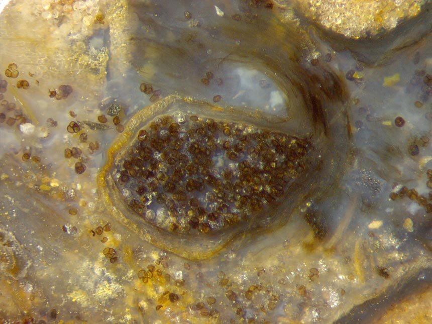
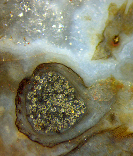
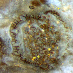
Fig.1:
4 Nothia
sporangia: with pale wall and dark spores (in the middle), with
pale wall and pale spores (in the corner above left), with dark wall
and
pale spores (above and below right); scattered spores, also in tetrads.
Image size 3.5mm, same scale for Figs.1-3.
Fig.2: Nothia sporangium
like a feeding bowl with diamonds:
black-coated sporangium with clear
spores.
Fig.3: Small Nothia sporangium
with cross-sections of the typical big tubes in the wall tissue
on the left. Image size 1mm.
The scattered spores in Fig.1, partially seen in tetrads, seem to be
hollow, with light-brown translucent walls, as they are also
known from Horneophyton,
for example. The light brown is possibly the natural residue of the
decayed organic matter.
The conspicuously different whitish aspect of the spores in the other
sporangia seen at the edges of Fig.1 might be the result of bleaching
by oxidation of the organic compounds left over after decay. Apparently
the bleaching has also affected a larger part of the sporangium wall
in the middle so that it has become pale yellow.
By contrast, the sporangium wall in Fig.2 is not
bleached but covered with
a black coating, probably
of microbial origin,
while the spores are bright and largely transparent and glittering.
The openings for spore release are
not or not clearly seen here.
What looks like an
indent in the sporangium wall in Fig.2 above left is possibly
indicating an opening slot.
(See also Rhynie
Chert News 33,
170
.)
Samples:
Rh7/24.2, found in 2003: Fig.1;
Rh3/11.3 (0.075kg), found
in 1998: Fig.2; Rh15/58.2 (0.21kg), obtained
from Barron
in 2011: Fig.3; (weights
refer to the samples
before cutting).
H.-J.
Weiss
2021
[1] H.
Kerp, H. Hass, V. Mosbrugger: New data on Nothia aphylla,
in:
P.G. Gensel, D.
Edwards (eds.): Plants Invade the Land, N.Y. 2001.
 |
 |
176 |