Croftalania
venusta and other Devonian
microbes
As a well-known but often overlooked fact, some
procaryotes are able to unite into surprisingly complex structures,
considering their primitive organisation on the cellular level.
Impressive examples are provided by the more or less dense tufts of
this filamentous blue-green alga or cyanobacterium [1] occasionally
seen fossilized in the Rhynie chert. Microbes hitherto
undescribed may have been involved in the
formation of horizontal microbial layers also found in the same sample.
The below pictures are to complement those in [1]. Also they
are shown
here for the beauty of the various aspects of Croftalania venusta.
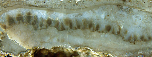

Figs.1,2: Croftalania
venusta
grown as separate tufts of filaments without
a solid substrate. Width of
Fig.1: 11mm.
Fig.3: Croftalania
grown on shrivelled Horneophyton.
Width
of the picture 10mm. 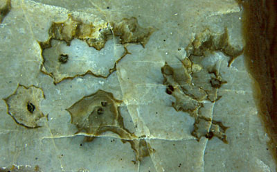
It is not obvious why the tufts in Figs.1,2 are connected by
what seems to be a wobbly dark line on the cut face, possibly the
boundary of some weakly
solidified or gel-like substrate. Croftalania
grows also on solid substrates (Figs.3-6), here
partially decayed Horneophyton
(which is easily mistaken for Nothia)
lying in water. It tends to grow upwards, towards the light.
There
are unexplained differences in the aspect which can be characterized in
a funny way by comparison with human hair: natural and fluffy
(Figs.1,2) or styled into spectacular and pointed shapes by means of
some sticky substance (Figs.3-6).
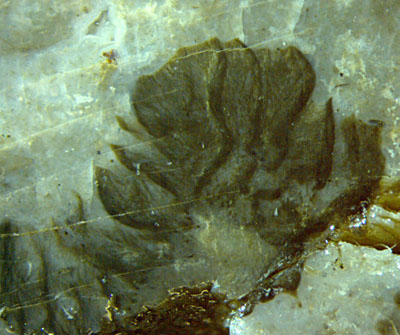
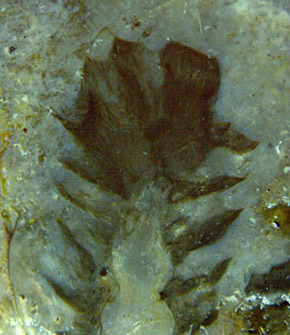
Figs.4,5: Pointed
tufts of Croftalania
with clear outline, resembling leaves or scales on a cone. Width of the
pictures 5.5mm, 4.3mm.
As
some of the pointed tips are seen to consist of converging filaments,
the idea suggests itself that they had been formed by capillary force
like the tip of a wet painter's brush. However, for this to happen the
habitat would have to temporarily fall dry, which most probably would
have irreversibly collapsed the delicate structures seen here.
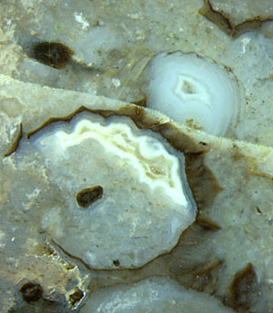
Fig.6: Cyanobacterial coating (Croftalania)
on Horneophyton
but not on Castracollis
moult fragment. Width of the picture 4.5mm.
Another feature requires an explanation: Distinct boundaries of
the filamentous tufts are more often present (Figs.3-6) than absent
(Figs.1,2). They look as if the tufts had been carefully trimmed with
smooth cuts regardless of the orientation of the filaments. Even plane
faces as in Fig.6 above right had been
formed in this way.
Note also the Castracollis
moult section. As none of the numerous moult fragments in this chert
sample is
coated with cyanobacteria, the little crustacean must have lived and
moulted in the water when the growth of Croftalania was
over. Hence, some time had been elapsed between the
growth of Croftalania
and the silicification of the flooded habitat, during which the
tufts could have been trimmed by means unknown, or possibly even by
crustaceans like Castracollis and
Ebullitiocaris,
smoothly nibbling off the organic gel with filaments enclosed.
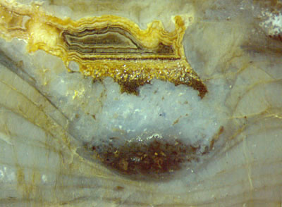
Fig.7: Microbial layers, originally horizontal,
largely deformed into a trough, and agate
indicating the absence of any further deformation while it was being
formed.
Width of the picture 4.5mm.
Stacks of more than a dozen horizontal layers up to several centimeters
in length, some of which are seen in Fig.7, have been observed
near Croftalania.
They resemble the continuous layers of doubtless microbial
origin in numerous other samples of Rhynie chert. In some places in the
present sample they are traversed by thin filaments which may be Croftalania, as
discussed in [1], where
also a possible involvement of other microbes is mentioned. Air bubbles
are
often seen trapped between the layers or having penetrated them when
ascending. The layers are always more or less deformed without a
discernable cause, as in the case of Fig.7, where 4 or 5 layers are cut
off, obviously in connection with the enigmatic formation of
a trough filled with dark debris and a water-filled cavity above, later
turned into agate. While the numerous layers of the banded agate were
deposited there
were no more deformations, as can be concluded from the perfectly
straight bottom line.
It
must be admitted that more questions than anwers are provided by the
above images of fossil cyanobacteria, which all have been taken from
one sample of Rhynie chert
found in 2001. Croftalania is
one of the very
few fossil cyanobacteria species [2] not confined to stromatolites.
Silicification of free water in the Devonian habitat of Rhynie had
enabled the leaf-like formations of Croftalania, which
anticipated the forms of eukaryotes to come, to be preserved as
life-like fossils.
H.-J.
Weiss
2013, emended 2014, 2015
[1] M. Krings, H. Kerp, H. Hass, T.N.
Taylor, N.
Dotzler:
A filamentous
cyanobacterium showing structured colonial growth from the Early
Devonian Rhynie chert.
Rev. Palaeobot. Palyn.
146(2007), 265-276.
[2] T.N. Taylor,
E.L. Taylor, M. Krings: Paleobotany,
Elsevier 2009, 115-117.
 |
 |
56 |