Pseudo-cells in Rhynie chert
 Occasionally,
cherts with or without silicified higher plants may show confusing
structures resembling plant tissue while being no such. In Fig.1, the
"cells" seen in the odd-shaped areas of decayed and vanished tissue in
a Nothia
rhizome do not seem to differ much from the
cells of the latter, at least with
respect to shape and size distribution. The conspicuous horizontal
boundary above the structure indicates the former presence of a watery
suspension.
Occasionally,
cherts with or without silicified higher plants may show confusing
structures resembling plant tissue while being no such. In Fig.1, the
"cells" seen in the odd-shaped areas of decayed and vanished tissue in
a Nothia
rhizome do not seem to differ much from the
cells of the latter, at least with
respect to shape and size distribution. The conspicuous horizontal
boundary above the structure indicates the former presence of a watery
suspension.
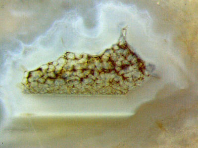
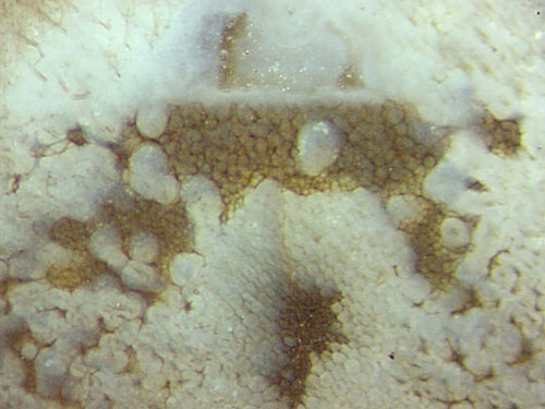
Fig.1 (right): Nothia rhizome
cross-section with central strand, surrounding tissue decayed and
replaced by coated fungus hyphae and pseudo-cellular structure grown in
formerly water-filled places.
Fig.2 (left): Formerly water-filled cavity with
thick lining of silica gel, grains of irregular shape grown inside.
What seems to be mutually contradictory, a
horizontal boundary above some structure in Fig.1 but
below in Fig.2, may be surprising. In Fig.2, a pale brown watery
suspension had settled at the bottom of a
water-filled cavity whose former contour can only be guessed, making
a horizontal surface, and eventually solidified into gel.
Then the remaining cavity got a
thin white lining all around, clearly seen only at the bottom where
it appears in
cross-section as a bright line. Then came a thick pale white lining, appearing
unevenly thick owing to varying orientation with respect to the cut
plane. The granular structure within does not look as much like tissue
as the structure in Fig.1 does. It seems that the horizontal boundary
of the latter, as well as a similar boundary in Fig.3, had been brought
about by some watery suspension settling in the lower part of the
cavity.
Fig.3 (below): Pseudo-cellular structures grown
in 3
formerly water-filled cavities between silica gel.
Width of the picture 4.3mm.
Figs.4,5,6: (below right): Details of Fig.3
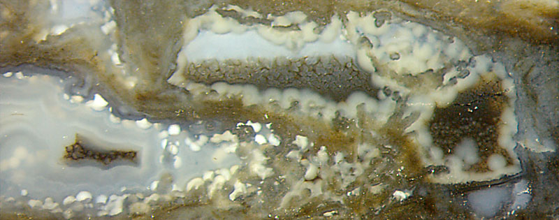
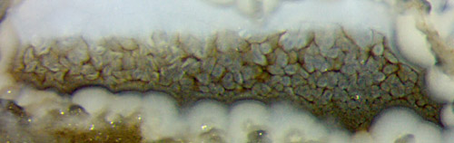
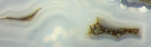
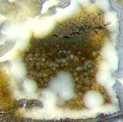
Fig.6 (right): Detail of Fig.3, possibly leading the way to
a partial explanation of the phenomenon of pseudocells.
The
pseudo-cellular structure is seen in Fig.6 above and below. Possibly
it originated from the abundant whitish clots. The growing globular clots would not fuse when
touching but keep their individuality and mutually squeeze into a
pattern resembling plant tissue. The illusion is favoured by
the
fact that the pseudo-cells are separated by dark boundaries which
possibly consist of microbes living on the surface of the clots or
pushed ahead by the growing clots and getting trapped.
Microbes
could even be the primary cause of the phenomenon. This is suggested by
the observation that the pseudo-tissue is often much darker than its
surroundings. On the left of Fig.5 there is a dark fill of
a narrow former cavity between whitish silica gel (all chalcedony now),
with only one small globular clot, apparently retarded in growth
compared to the bigger clots on the right of Fig.5. Hence, one may
assume that first came the dark, then the clots.
Dark deposits, layers, and crusts occasionally seen in the Rhynie chert are
most probably due to microbial activity (See Rhynie Chert News 87 .) If looked at superficially, pseudo-tissue produced by microbes could be confused with real tissue.
H.-J.
Weiss 2017 2020
 |
 |
112 |