Dainty meshwork aspect of Aglaophyton
epidermis
 The
Lower Devonian plant Aglaophyton
( = Rhynia major),
which is abundantly seen in the Rhynie chert (Fig.1), does
not often show a distinct view of its epidermis. The rare cases where
it does may look surprisingly different as a result of not yet
explained peculiarities of fossilisation.
The
Lower Devonian plant Aglaophyton
( = Rhynia major),
which is abundantly seen in the Rhynie chert (Fig.1), does
not often show a distinct view of its epidermis. The rare cases where
it does may look surprisingly different as a result of not yet
explained peculiarities of fossilisation.
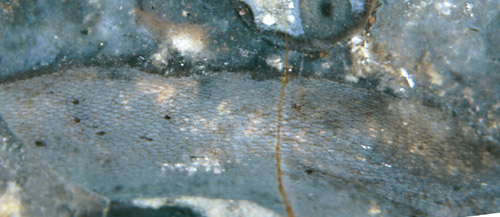
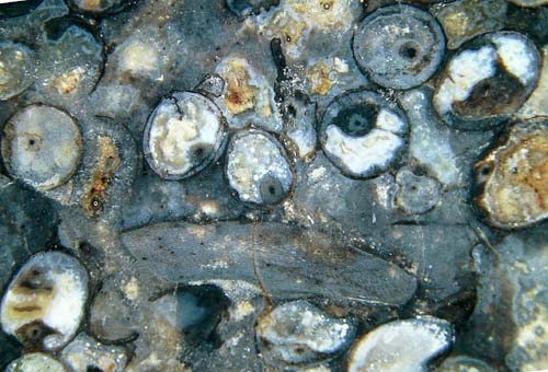
Fig.1 (right): Aglaophyton
sections in Rhynie
chert, in various states of decay; one fragment seen in lateral view,
with uncommonly well
preserved epidermis pattern. See enlarged details. Width of the
picture 20mm.
Fig.2 (left): Aglaophyton
epidermis
with unevenly distributed stomata, detail of
Fig.1. Width of the picture 8mm.
Fig.3 (below left): Aglaophyton
epidermis,
detail of Fig.1, width of the picture 3mm.
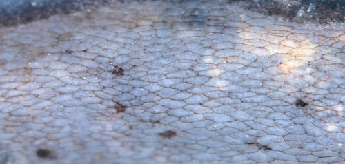
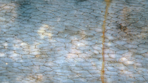
Fig.4 (at the bottom): Aglaophyton
epidermis, detail of Fig.1, only one stoma seen here,
width of the picture 2.3mm.
The unexpectedly distinct surface pattern is apparently
brought about by tiny grooves filled with cuticle substance between
adjacent epidermis cells. The meshwork appears even more delicate in
Fig.4. Cell edges beneath the surface are faintly seen through the
transparent
chalcedony on a few cells in Figs.3,4.
A quite different and even more enigmatic aspect of the epidermis of Aglaophyton
is shown in Rhynie Chert News 6,
where there is a conspicuous polygonal black frame with
fitting shape placed right below the upper face of every epidermis
cell.
H.-J. Weiss
2012
 |
 |
47 |