A rare sight of Ventarura
tissues (1)
 Among
the early land plants in the Lower Devonian Rhynie chert, Ventarura
is
distinguished by a peculiarity: rapid decay of tissues
except for a fraction of cortex (Fig.1). The latter is often
but not
always seen on cross-sections of aerial axes as
a surprisingly well-preserved enigmatic ring of cells but the epidermis
has apparently not been seen hitherto, as can be concluded from
quotations like "... cells directly internal to the cuticle are not
preserved" and "... an unequivocal epidermal layer was not observed
..." [1]. This is confirmed by "... an epidermal layer has not been
seen ..." [2]. In view of the fact that the epidermis
is seen on all
other land plants in the Rhynie chert, often very clearly, one could
suspect that its absence in the silicified aerial axes of Ventarura
might be
restricted to the one large chert pod which the publication [1] is
based on. Judging from several hundred Ventarura
sections seen on more than a dozen own chert samples which apparently
represent a
wider variety of fossilisation conditions it can be stated that the epidermis
is really almost never seen on Ventarura
aerial axes. One such is
seen in Fig.2. By lucky incidence, this specimen offers sections of
epidermis cells in three mutually perpendicular planes.
Among
the early land plants in the Lower Devonian Rhynie chert, Ventarura
is
distinguished by a peculiarity: rapid decay of tissues
except for a fraction of cortex (Fig.1). The latter is often
but not
always seen on cross-sections of aerial axes as
a surprisingly well-preserved enigmatic ring of cells but the epidermis
has apparently not been seen hitherto, as can be concluded from
quotations like "... cells directly internal to the cuticle are not
preserved" and "... an unequivocal epidermal layer was not observed
..." [1]. This is confirmed by "... an epidermal layer has not been
seen ..." [2]. In view of the fact that the epidermis
is seen on all
other land plants in the Rhynie chert, often very clearly, one could
suspect that its absence in the silicified aerial axes of Ventarura
might be
restricted to the one large chert pod which the publication [1] is
based on. Judging from several hundred Ventarura
sections seen on more than a dozen own chert samples which apparently
represent a
wider variety of fossilisation conditions it can be stated that the epidermis
is really almost never seen on Ventarura
aerial axes. One such is
seen in Fig.2. By lucky incidence, this specimen offers sections of
epidermis cells in three mutually perpendicular planes.
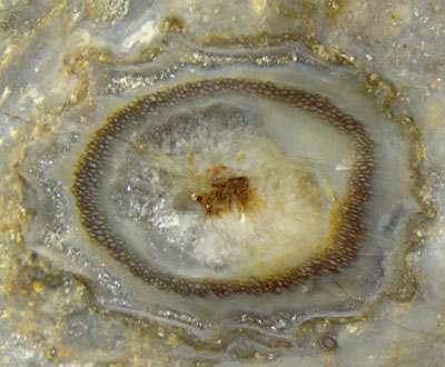
Fig.1 (above):
Ventarura
cross-section, typical aspect: well-preserved ring-shaped
fraction of cortex, shrivelled surface, quartz-filled cavities
replacing decayed and vanished tissue. (There is no central strand
left. The brown spot is a deposit of iron ore between quartz.)
Width of the picture 6mm.
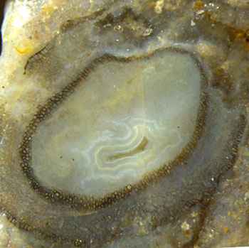 Fig.2: Ventarura
cross-section on the raw chert surface, unique aspect with patches of
epidermis and faintly seen outer cortex in addition to the
conspicuous mid-cortex, no inner cortex and central
strand left.
Fig.2: Ventarura
cross-section on the raw chert surface, unique aspect with patches of
epidermis and faintly seen outer cortex in addition to the
conspicuous mid-cortex, no inner cortex and central
strand left.
Large diagonal of the section 6mm. Same chert sample as Fig.1.
The unique cross-section
in Fig.2 with patches of tissue other than the characteristic ring
in mid-cortex has been found on the raw surface of a small sample of
38g among sections of "normal" aspect as seen also on the raw surface
and on the cut and polished
faces (Fig.1).
The
enlarged view in Fig.3 shows clearly the epidermis at the bottom of the
picture, slightly wavy owing to slight degradation and shrinkage of the
outer cortex.
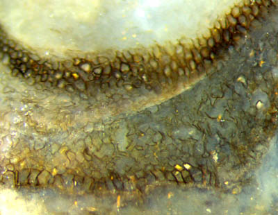
Fig.3 (right): Ventarura
tissue layers: epidermis, slightly
degraded outer cortex, well preserved
mid-cortex, vanished inner cortex. Detail of Fig.2, width of the
picture 2mm.
There
is no indication of a reason why only in this particular case some
patches of the
epidermis are in a similar state of good preservation as the
mid-cortex,
the latter being an enigma in itself. From the aspect of the slightly
degraded outer cortex
it can be concluded that
cell sizes and their variation are essentially the same as in
mid-cortex. These observations are confirmed by Fig.4.
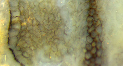 Fig.4 (left): Ventarura
tissue layers, left to right: cuticle with black deposit,
epidermis, slightly degraded outer
cortex, well preserved mid-cortex, vanished inner cortex.
Fig.4 (left): Ventarura
tissue layers, left to right: cuticle with black deposit,
epidermis, slightly degraded outer
cortex, well preserved mid-cortex, vanished inner cortex.
Detail of Fig.2, width of the picture 1.3mm.
As a lucky incidence, the shoot seen in
cross-section near the edge of one fracture face (Fig.2)
appears in approximately lengthwise section beyond that edge, with a
very small strip of epidermis seen in top view (Fig.5).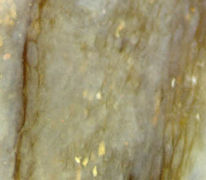
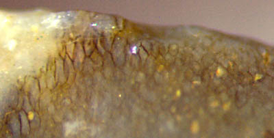
Fig.5: Ventarura
seen from outside, epidermis seen as a small strip of lenghty
cells on the left, broken off elsewhere so
that the below cortex
is seen there.
Fig.6 (far right): Ventarura
tissue layers, approximately lengthwise section,
left to right: epidermis, degraded
outer cortex, mid-cortex.
Since the elusive epidermis of Ventarura
has apparently never been seen and described, the small patches of
epidermis seen here in cross-section (Figs.3,4), lengthwise section
(Fig.6), and top view (Fig.5) fill a gap in the knowledge of this still
enigmatic plant. (See Part 2.)
H.-J.
Weiss
2014
[1] C.L.
Powell, D.
Edwards, N.H. Trewin: A new vascular plant from the
Lower Devonian Windyfield chert, Rhynie, NE Scotland.
Trans. Roy. Soc. Edinburgh, Earth Sci.
90(2000 for 1999), 331-349.
[2] D.
Edwards: Embryophytic sporophytes in the Rhynie
and Windyfield cherts.
Trans. Roy. Soc. Edinburgh,
Earth Sci.
94(2004 for 2003), 397-410.
 |
 |
61 |