 Trichopherophyton
sporangia
Trichopherophyton
sporangia
Among the early land plants found in the Rhynie
chert, Trichopherophyton
is distinguished by pointed bristles on its upper parts, meant
to deter
herbivores (Fig.1). Fragmentary sporangia contours seen on
chert samples (Fig.2) are easily confused
with those of Ventarura
[1] or
Asteroxylon
[2] unless some of the sparsely distributed bristles are seen attached
or nearby.
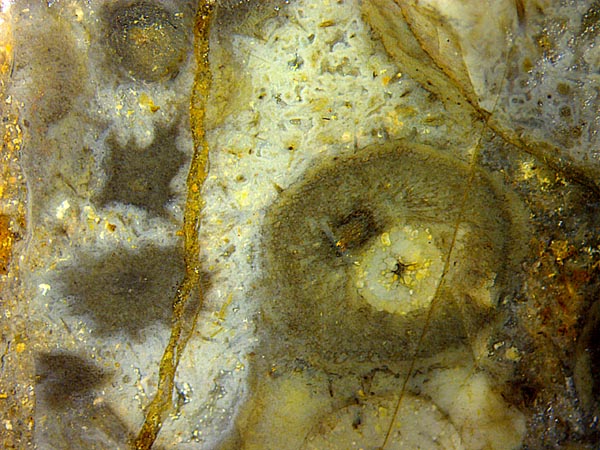
Fig.1 (right):
Trichopherophyton
cross-sections
with bristles, shrivelled ones on the left; fungus hyphae coated with
white chalcedony. Image width 7mm.
Remarkably, Trichopherophyton
sporangia contours are seen in Figs.2,3 on two mutually perpendicular fracture
faces forming an edge on this chert sample. Note that the
filled sporangium in Fig.3 is the same as the left one in Fig.2, and
the empty sporangium in Fig.3 is the same as the middle one in Fig.2.
Blurred details in Fig.2 are due to uneven face.
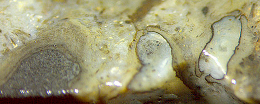 Fig.2 (right): Raw sample
edge with Trichopherophyton
sporangia with open slots; vaguely seen bristles mostly detached.
Fig.2 (right): Raw sample
edge with Trichopherophyton
sporangia with open slots; vaguely seen bristles mostly detached.
Fig.3 (below far right): Same
sample edge as in Fig.2, view on the face beyond the edge,
shifted to the right.
Image width 5mm. Same scale for Figs.2-6.
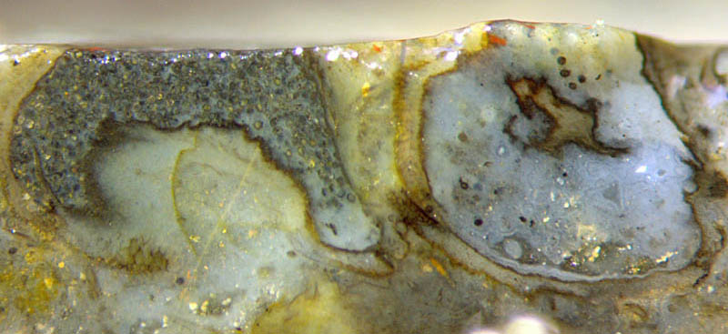
What
is hidden below the mass of spores in Fig.2 is seen in Fig.3: It is a
broad pale part of sporangium wall surrounding a likewise pale pad of tissue. Apparently the empty
sporangium with different orientation on
the right in Fig.3 shows the natural fracture face incidentally cutting through an
irregular boundary like the one seen in
the left sporangium.
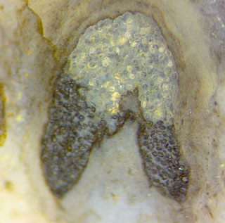
Fig.4 (right): Trichopherophyton
sporangium on the raw sample surface, same sample as Figs.1-3;
spores with differential stages of development
and preservation for reasons unknown.
The
dark spores seem to be not fully developed but affected with black
microbial coatings. Partially they are still arranged in rows.
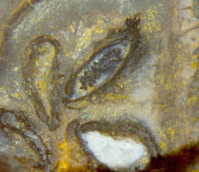
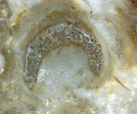
Fig.5 (far left): Cluster of sporangia with open
slots; Trichopherophyton
bristles on the right.
Fig.6:Trichopherophyton
sporangium with bristle.
The 3-D shape of Trichopherophyton
sporangia is not easily derived from the
sections in Figs.2-6.
Possibly a model drawing
for Asteroxylon
sporangia in [2] comes
close to this case, too.
Samples:
Rh4/74 (0.17kg) found in 2000: Figs.1-4.
Rh15/78.2 (0.58kg) from Barron
2004: Fig.5.
Rh2/71.3 (3.22kg)
found in 2001: Fig.6.
H.-J.
Weiss
2021
[1] www.abdn.ac.uk/rhynie/venta.htm
[2] H. Kerp, C.H.
Wellmann,
M. Krings, P. Kearney, H. Hass:
Reproductive organs and
in-situ spores of Asteroxylon
... Int. J. Plant Sci. 174(3), 293-308, 2013.
 |
 |
175 |